
Retinal angiography
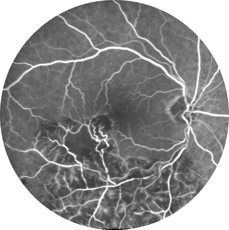
Photographs of the back of the eye after injection of a fluorescent dye, to show precisely the condition of the retinal blood vessels — at the practice, during a dedicated consultation, by Dr Srour and Dr Chetrit.
Retinal angiography: the essentials in 30 seconds
- Photographs the circulation of the retina using a fluorescent dye injected into a vein in the arm.
- Essential in some forms of AMD, in diabetic retinopathy and in retinal vein occlusions.
- Your skin and urine may be coloured for a few hours: this is an expected and harmless effect. Tell us before the test about any known allergy.
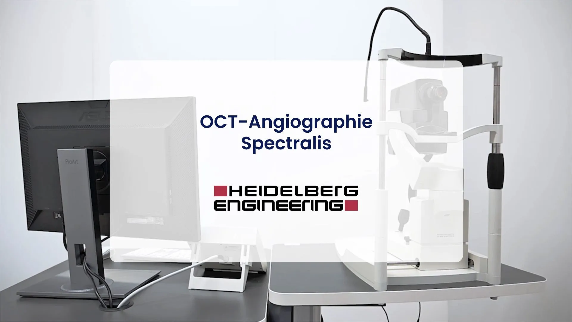
- Performed at the practice; OCT angiography, which needs no injection, replaces it in many cases.
What is retinal angiography?
Thanks to advances in OCT and OCT angiography (no injection), angiography is now prescribed in a more targeted way — but it remains indispensable in certain situations: it alone shows the dynamics of circulation (leakage, non-perfused areas) across the whole retina.
The test consists of taking photographs of the back of the eye after injecting a fluorescent dye — fluorescein or indocyanine green — into a vein in the arm. As it circulates through the eye, the dye reveals the condition of the blood vessels of the retina and the choroid (the layer of blood vessels behind the retina): leakage, occlusions, poorly perfused areas, abnormal vessels.
It complements OCT angiography (no injection) when a dynamic or deep analysis is needed, and is used for the diagnosis, monitoring and follow-up of retinal conditions treated with anti-VEGF injections (intravitreal injections, IVT).
When is it indicated?
What happens during the examination?

Precautions & side effects: what you need to know
Angiography is a very well tolerated test, preceded by a medical questionnaire: tell us about your allergies (in particular to dyes or iodine), your history and your current treatments — premedication may be offered if necessary.
Please also tell us if you are pregnant or breastfeeding: the indication is then reassessed with your doctor, and the test postponed if it is not essential.
Download the SFO leaflet — Fluorescein angiography (PDF)Retinal angiography in Noisy-le-Grand
The Grand Paris Ophtalmologie practice performs retinal angiography on site, during a dedicated consultation, for patients from Noisy-le-Grand and eastern Paris (93, 94, 77): Bry-sur-Marne, Villiers-sur-Marne, Champs-sur-Marne, Chelles, Torcy and Marne-la-Vallée, as well as Le Perreux-sur-Marne, Nogent-sur-Marne, Fontenay-sous-Bois, Vincennes, Saint-Maur-des-Fossés, Créteil, Montreuil and Rosny-sous-Bois — a test usually reserved for specialist centres, without having to go to Paris, interpreted by Dr Mayer Srour, a retina specialist, and Dr Yaïr Chetrit.
Angiography: your questions answered
Answers from Dr Mayer Srour and Dr Yaïr Chetrit, ophthalmologists in Noisy-le-Grand.
Will my skin and urine be yellow after the test?
Yes, with fluorescein: this is normal and harmless — the dye is naturally eliminated within 24 to 48 hours. Drink plenty of fluids after the test to speed up elimination.
Is the test painful?
No — only the placement of the venous line, which is equivalent to a blood test, and the flash of the photographs is briefly dazzling. A feeling of warmth or slight nausea after the injection is possible, brief and harmless.
Can I drive afterwards?
No: your pupils are dilated and your vision remains blurred and dazzled for 2 to 3 hours. Come accompanied or by public transport.
Do I need to fast?
No, you do not need to fast. However, do remember to bring your medical file and the list of your treatments for the pre-test questionnaire.
Are there any risks or side effects?
The test takes place without side effects in the vast majority of cases. Your skin and urine may be coloured yellow for about 24 hours. A questionnaire checks for any history of allergy; severe reactions are exceptional.
What is the difference from OCT angiography?
OCT angiography shows the vessels without an injection, in a few seconds — it is the first-line test. Angiography with dye remains essential to see leakage in real time, to explore the deep choroid (indocyanine) or to assess peripheral ischaemia. Both are available at the practice.
How much time should I allow?
The test lasts ≈ 10 minutes (fluorescein) to 30 minutes (indocyanine), but allow more time for dilation and the questionnaire: about 1 hour to 1 hour 30 minutes at the practice.
Related reading
Have you been prescribed an angiogram?
The test is performed at the Noisy-le-Grand practice, during a dedicated consultation.
The Grand Paris Ophtalmologie practice in Noisy-le-Grand provides consultations, screening and surgery for patients from neighbouring towns: