
The OCT angiography module: retinal vessels, without injection
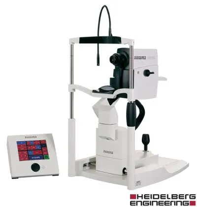
Mounted on our Spectralis, the OCT angiography (OCT-A) module maps the blood flow of the retina without injecting dye, whereas conventional angiography requires a fluorescein infusion.
What is OCT angiography, and what is it used for?
OCT-A is an evolution of OCT: by repeating scans very quickly at the same spot, the device detects what moves (the red blood cells in the vessels) and thus reconstructs a map of retinal circulation, without injecting any product.
It visualises the vascular networks layer by layer (superficial plexus, deep plexus, choriocapillaris): from the neovessels of AMD to the ischaemic areas of diabetes, the vasculature can be read directly on screen, within seconds.
Why it is a benchmark in its category
Fluorescein angiography remains a valuable test, but it requires a dye infusion, takes about thirty minutes, colours the skin and urine, and carries rare allergic risks. OCT-A obtains a vascular map without any of these drawbacks, so it can be repeated at every visit.
Backed by the Spectralis eye-tracking, the module overlays vascular maps from one visit to the next, ideal for monitoring a neovessel treated by intravitreal injections. Dr Mayer Srour has also published several scientific studies on OCT-A in international journals.
When the practice uses it
What it means for you
OCT angiography: your questions
Answers from Dr Yaïr Chetrit and Dr Mayer Srour, ophthalmologists in Noisy-le-Grand.
Does OCT-A replace fluorescein angiography?
In many follow-up situations, yes. But dye angiography remains useful for certain diagnoses (leakage, periphery): your doctor chooses the appropriate test, and both are carried out at the practice.
Does the test carry any risk?
None: no injection, no dye, no X-rays, just light. It can be repeated without limit, including in pregnant women or in cases of allergy.
Is any special preparation needed?
No. The test is carried out like a standard OCT, most often without dilation: you leave as normal.
Related reading
Need vascular monitoring of the retina?
The OCT-A module is built into the Spectralis at the Noisy-le-Grand practice: the test is done on site, without injection.
The Grand Paris Ophtalmologie practice in Noisy-le-Grand provides consultations, screening and surgery for patients from neighbouring towns: