

Ocular ultrasound (eye ultrasound scan): B-scan & UBM
The B-scan examines the back of the eye (retina, vitreous); the UBM analyses the front of the eye (anterior segment) at very high resolution.
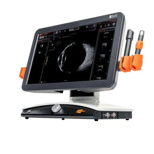
Eye ultrasound: the essentials in 30 seconds
- B-scan: ultrasound through the closed eyelid to see the retina and vitreous when the back of the eye cannot be seen (dense cataract, haemorrhage).
- UBM (ultrasound biomicroscopy): high-frequency ultrasound of the anterior segment: drainage angle, iris, ciliary body. Essential for certain types of glaucoma, iris cysts and tumours, and for assessment before an implant.
- Where can I have a UBM ultrasound? At the practice in Noisy-le-Grand, on your ophthalmologist's prescription or as part of your follow-up — with no hospital visit.
- What to expect: 10 to 15 minutes, painless; for the UBM, one drop of anaesthetic and a small water cup placed on the eye. You can drive afterwards.
What is ocular ultrasound?
Ocular ultrasound uses ultrasound waves — sound that cannot be heard — to explore the inside of the eye. It is the same principle as the ultrasound used elsewhere in medicine: no X-rays, no injection. The probe emits waves that bounce off the structures of the eye, and the machine builds an image.
Its great advantage: it can “see” the eye even when it is not transparent — when a very dense cataract or bleeding in the vitreous prevents a view of the back of the eye. Two complementary techniques exist: the B-scan and the UBM.
B-scan or UBM?
Two ultrasound techniques, two areas of the eye explored — they are complementary:
When is it useful?
What an eye ultrasound shows
B-scan ultrasound explores everything that a fundus exam cannot show when the media are opaque: the vitreous (haemorrhage, inflammation, posterior detachment), the retina (tear, detachment), the choroid, the optic nerve head and any intraocular masses. The very high-frequency UBM analyses the anterior segment: drainage angle, iris, ciliary body and the position of an implant. The report is given and explained on the same day.
UBM ultrasound in practice: indications, what to expect, cost
The UBM (ultrasound biomicroscopy) is a very high-frequency ultrasound scan (35 to 50 MHz) that produces a cross-sectional image, down to a tenth of a millimetre, of the front of the eye: cornea, drainage angle, iris, ciliary body and lens. It is the only examination that shows what lies behind the iris, where OCT and the slit lamp cannot see. The practice is equipped with it and performs it on site, in Noisy-le-Grand.
What to expect: one anaesthetic drop, then a small cup filled with fluid is placed between the eyelids; the probe does not touch the cornea. The examination lasts 10 to 15 minutes for both eyes, without dilating the pupil, and you can drive afterwards. To study the angle, the examination is carried out in dim light and then in light, to see how the angle behaves.
Where can I have a UBM ultrasound? Few practices in Île-de-France are equipped with it: ours sees patients referred by their ophthalmologist (93, 94, 77, east Paris) as well as those followed at the practice. The report, with images, is given on the same day and sent to the referring doctor. No prescription is needed, but a letter from your ophthalmologist stating the question to be answered is useful.
Cost: €130 to €160 depending on the extent of the examination (sector 2 OPTAM: doctors who may charge above the standard rate, within a capped scheme); the French national health insurance (Sécurité sociale) reimbursement base is €52.80, and the balance is generally covered by your top-up private health insurance (mutuelle). Details on the fees and reimbursement page.
What happens during the examination?
Nothing could be simpler, and it is completely painless. For the B-scan, a little gel is applied and the probe is gently placed on the closed eyelid; you are asked to move your eye in different directions. For the UBM, an anaesthetic drop is instilled because the probe explores the front of the eye. The examination takes a few minutes, with no X-rays or injection, and the images are interpreted immediately by your ophthalmologist.
No special preparation: no fasting, no need to stop any treatment. It is simply advised to remove your contact lenses before the examination.
Ocular ultrasound in Noisy-le-Grand
The Grand Paris Ophtalmologie practice performs B-scan and UBM ultrasound on site, for patients from Noisy-le-Grand and east Paris (93, 94, 77): Bry-sur-Marne, Villiers-sur-Marne, Champs-sur-Marne, Chelles, Torcy and Marne-la-Vallée, as well as Le Perreux-sur-Marne, Nogent-sur-Marne, Fontenay-sous-Bois, Vincennes, Saint-Maur-des-Fossés, Créteil, Montreuil and Rosny-sous-Bois. The examination is carried out and interpreted by Dr Yaïr Chetrit or Dr Mayer Srour.
Ocular ultrasound: your questions
Answers from Dr Yaïr Chetrit and Dr Mayer Srour, ophthalmologists in Noisy-le-Grand.
Is a UBM eye ultrasound painful?
No, the examination is painless. For the B-scan, the probe is placed on the closed eyelid with a little gel; for the UBM, an anaesthetic drop is instilled. No X-rays, no injection.
B-scan or UBM of the eye: what is the difference?
The B-scan explores the back of the eye (retina, vitreous); the very high-frequency UBM analyses the anterior segment (cornea, iris, angle, ciliary body). The two are complementary.
What happens during an eye ultrasound?
Nothing could be simpler, and it is painless. For the B-scan, a little gel is applied and the probe is gently placed on the closed eyelid; for the UBM, an anaesthetic drop is instilled and then a small cup filled with fluid is placed between the eyelids. The UBM takes 10 to 15 minutes, without dilating the pupil, and you can drive afterwards.
How much does a UBM ultrasound cost?
€130 to €160 depending on the extent of the examination, in sector 2 OPTAM (doctors who may charge above the standard rate, within a capped scheme). The French national health insurance (Sécurité sociale) reimbursement base is €52.80; the balance is generally covered by your top-up private health insurance (mutuelle). The amount is stated before the examination.
Do I need a prescription for a UBM?
No: you can see an ophthalmologist directly. A letter from your ophthalmologist stating the question to be answered (angle, cyst, implant) is, however, useful for targeting the examination.
When is a B-scan essential?
Mainly when the back of the eye cannot be seen (very dense cataract, vitreous haemorrhage), to look for a retinal detachment or a tumour.
What is the UBM used for?
To explore the angle (narrow-angle glaucoma), tumours and cysts of the iris or ciliary body, and to check the position of an implant.
Where can I have a UBM ultrasound in Île-de-France?
Few community-based practices have a UBM machine. The Grand Paris Ophtalmologie practice in Noisy-le-Grand (93) performs UBM and B-scan on site, for its own patients as well as those referred by a colleague, with the report given on the same day. Appointments by telephone or on Doctolib.
Is an eye ultrasound reimbursed?
Yes: B-scan and UBM are listed procedures, covered by the French national health insurance (Assurance Maladie) when medically justified. The fees charged are shown on the Fees and reimbursement page.
Related reading
Has an ocular ultrasound been prescribed for you?
The examination is carried out at the Noisy-le-Grand practice and interpreted immediately.
The Grand Paris Ophtalmologie practice in Noisy-le-Grand provides consultations, screening and surgery for patients from neighbouring towns: