
Myopia
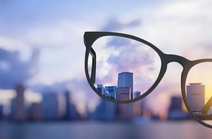
Myopia makes distance vision blurred: it can be corrected with glasses, contact lenses or surgery. But beyond a certain degree — high myopia — the eye becomes too long and the retina becomes more fragile: it is then a genuine condition requiring lifelong monitoring. High myopia is a speciality of Dr Mayer Srour, who provides screening, imaging and treatment on site in Noisy-le-Grand together with Dr Yaïr Chetrit.
High myopia: the essentials in 30 seconds
- High myopia = beyond −6 dioptres or a very long eye: the stretched retina is more fragile.
- Risks to screen for: retinal tears and detachment, abnormal new vessels in high myopia, and myopic maculopathy.
- A regular dilated fundus examination (at least once a year) is recommended, even without symptoms.
- Flashes, a shower of floaters or distortion: seek urgent consultation.
What is myopia?
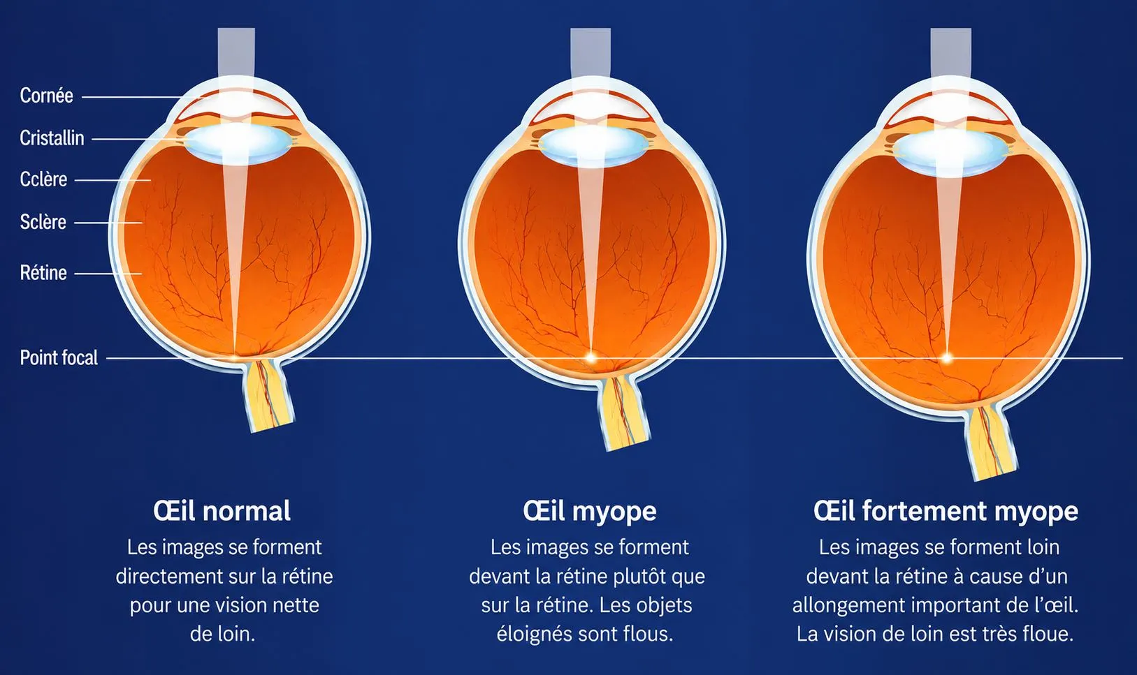
Myopia is not a disease but a very common refractive error: the eye is slightly too long, or its cornea too curved, so that the image forms in front of the retina. As a result, distance vision is blurred, while near vision remains sharp. It most often appears in childhood or adolescence, progresses until the age of 20 to 25, then stabilises.
Myopia is measured in dioptres (a figure preceded by a “−” sign). It is corrected with glasses, contact lenses or refractive surgery (laser or implant) once vision has stabilised.
What is high myopia?
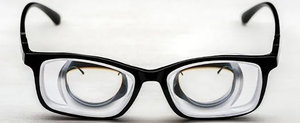
A myopic eye is an eye that is too long: the image forms in front of the retina and distance vision is blurred. We speak of high myopia (or severe myopia) beyond −6 dioptres, or when the length of the eye exceeds 26 mm (compared with 23.5 mm for a normal eye).
At this degree, the elongation of the eyeball permanently stretches the retina, the choroid and the wall of the eye. This stretching weakens the tissues and exposes the eye, throughout life, to complications that can threaten vision. High myopia is therefore both an optical error and a retinal condition requiring monitoring.
Two situations to distinguish
Not all people with high myopia are alike. The distinction is essential: it determines how often monitoring is needed and the risk to vision.
The shift from one to the other happens gradually, sometimes after several decades. This is the whole value of regular OCT monitoring: spotting the first signs of degeneration before they affect vision.
Complications & warning signs
Because the eye is too long, high myopia increases the risk of several eye diseases. Most can be treated successfully if detected early — hence the importance of monitoring.
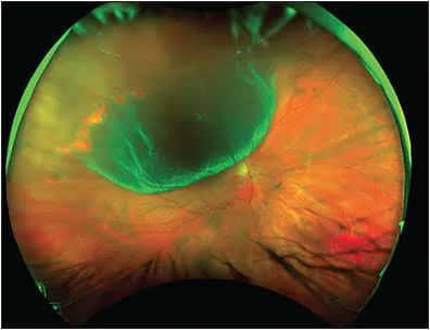
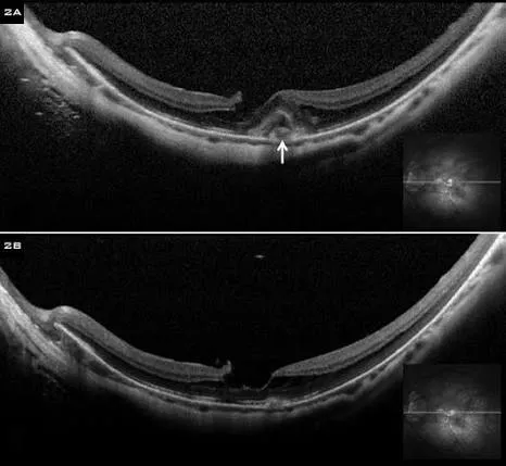
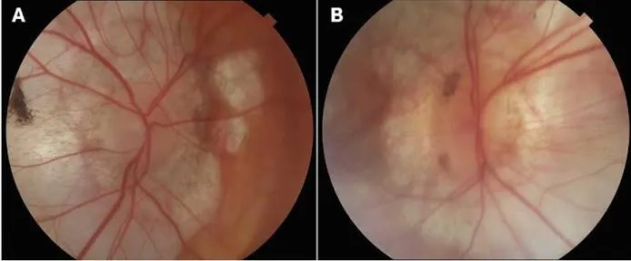
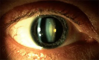
See a doctor urgently for: a shower of floaters, flashes of light, a veil or shadow in the visual field, or straight lines that appear distorted. These signs may herald a tear, a retinal detachment or abnormal new vessels — the practice sees emergencies on the same day.
Screening and monitoring at the practice
Monitoring in high myopia relies on retinal imaging. Everything is carried out in a single visit on the practice’s technical platform:
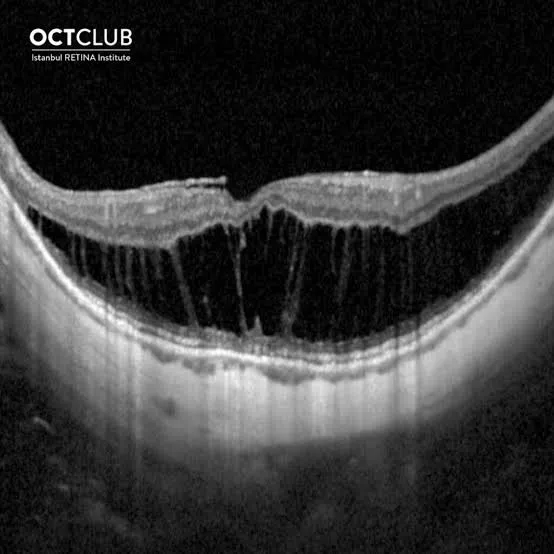
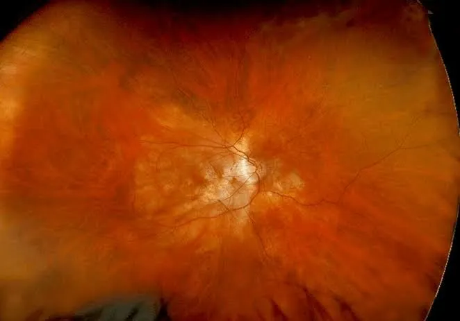
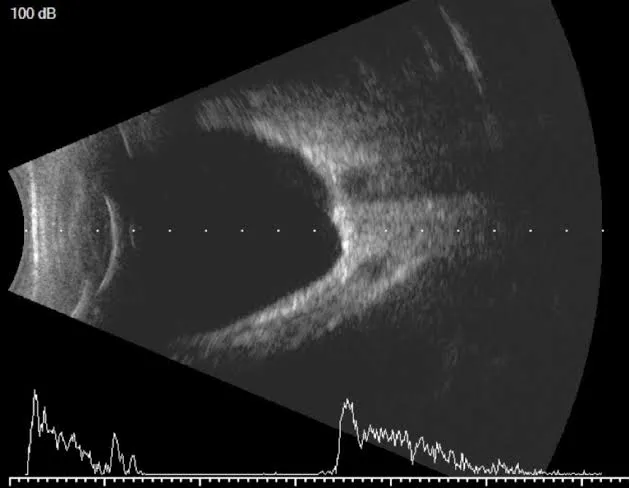
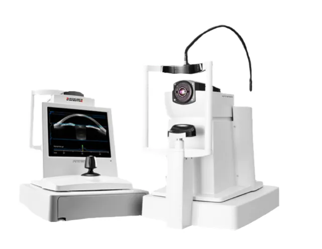
Recommended frequency: one complete retinal assessment per year, with a dilated fundus examination. It is brought forward (every 6 months) in cases of very high myopia, staphyloma or a history of complications.
Correcting vision, treating complications
Management is twofold: correcting the refractive error and treating complications if they occur.
When myopia appears early, it is now possible to slow its progression to limit the risk of high myopia in adulthood: low-dose atropine eye drops or myopia-control lenses — combined with time spent outdoors and limited screen time.
Important: correcting vision (glasses, laser, implant) does not remove the retinal risk linked to the length of the eye. Retinal monitoring remains essential, even after surgery.
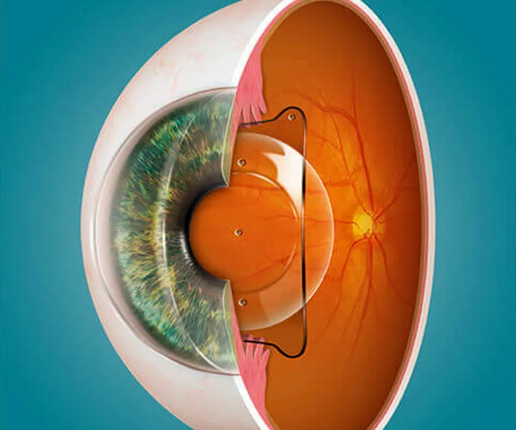
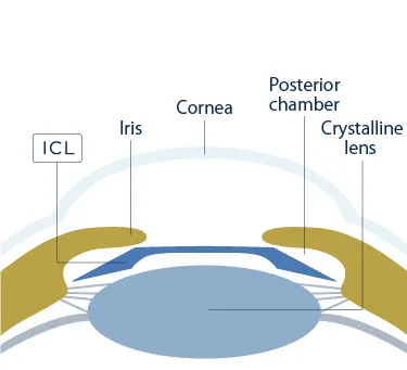
When myopia is too high for laser (beyond about −8 to −10 D) or the cornea is too thin, the phakic implant is the reference solution. It is a thin, flexible lens slipped inside the eye, in front of the natural lens, without touching the cornea.
The indication is made after a full assessment (corneal topography, biometry, corneal cell count). See also refractive surgery.
High myopia: good habits
Even without symptoms and even when well corrected: the annual fundus examination detects complications before they affect vision.
Flashes, floaters, a veil, distorted lines: these signs call for urgent consultation — the practice sees patients on the same day.
Be careful with contact sports and blows to the eye, which increase the risk of retinal detachment in high myopia.
Childhood myopia can now be slowed: early screening and monitoring limit the risk of high myopia in adulthood.
Official factsheets from the Société Française d'Ophtalmologie
Written by SFO experts, they describe how each treatment is carried out, and its benefits and risks. Please read them before your consultation.
High myopia in Noisy-le-Grand
The Grand Paris Ophtalmologie practice provides screening, imaging and monitoring of high myopia for patients from Noisy-le-Grand and eastern Paris (93, 94, 77): Bry-sur-Marne, Villiers-sur-Marne, Champs-sur-Marne, Neuilly-sur-Marne, Gournay-sur-Marne, Noisiel, Torcy, Chelles, Gagny, Le Perreux-sur-Marne, Nogent-sur-Marne, Créteil and Marne-la-Vallée — OCT, wide-field retinal photography and treatments on site.
Dr Mayer Srour, trained at the CHI de Créteil and a specialist in the retina and macula, and Dr Yaïr Chetrit provide monitoring for high myopia in liaison with your optician and your GP (médecin traitant).
Good to know: in cases of significant visual loss due to pathological myopia, low vision rehabilitation and optical aids can be offered to preserve independence in daily life.
High myopia: your questions
Answers from Dr Mayer Srour and Dr Yaïr Chetrit, ophthalmologists in Noisy-le-Grand.
What is high myopia?
Myopia beyond −6 dioptres, or an eye that is too long (more than 26 mm). The stretched eye weakens the retina: high myopia is both a refractive error and a retinal condition requiring monitoring.
Is high myopia dangerous?
It increases the risk of retinal detachment, maculopathy and abnormal new vessels, glaucoma and early cataract. Most can be treated successfully if detected early.
How often should it be monitored?
Generally once a year: dilated fundus examination, macular OCT and wide-field retinal photography. The frequency is increased if myopia is very high or if there is a history of complications.
Floaters, flashes: is it serious?
These signs should be taken all the more seriously in high myopia. A shower of floaters, repeated flashes or a veil call for urgent consultation so that the retina can be examined.
Can high myopia be operated on?
Yes: laser for myopia up to about −8 D, implant (ICL) beyond that. But correcting vision does not remove the retinal risk: monitoring remains essential, even after surgery.
Is high myopia hereditary?
There is a family predisposition. In children, the progression of myopia can now be slowed (low-dose atropine, myopia-control lenses).
Which tests are used to monitor the retina?
At the practice: OCT, wide-field retinal photography, biometry (length of the eye) and B-scan ultrasound if needed.
Does myopia stabilise with age?
Low and moderate myopia stabilise at around 20 to 25 years. High myopia, however, may continue to progress slowly throughout life — hence the value of regular monitoring.
Are monitoring and treatments reimbursed?
Yes: the consultation, retinal imaging (OCT, retinal photography), laser and injections are covered by the Assurance Maladie (the French national health insurance). Elective refractive surgery, however, is not reimbursed.
Related reading
Highly myopic? Have your retina monitored.
Screening and monitoring of high myopia at the Noisy-le-Grand practice — on-site retinal imaging, emergencies seen on the same day.
The Grand Paris Ophtalmologie practice in Noisy-le-Grand provides consultations, screening and surgery for patients from neighbouring towns: