

Keratoconus
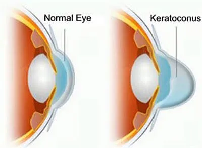
Keratoconus is a progressive deformation of the cornea, which thins and takes on a cone shape. It often appears in adolescence and blurs vision. Detected early by topography, it can be stabilised and vision corrected.
Keratoconus: the essentials in 30 seconds
- Progressive deformation of the cornea, most often starting in adolescence; rubbing the eyes makes it worse.
- Warning signs: astigmatism that changes quickly, double vision, a correction that is never satisfactory.
- Cross-linking halts progression; rigid or scleral lenses for vision.
- Screening by corneal topography at the practice — also essential before any refractive surgery.
What is keratoconus?
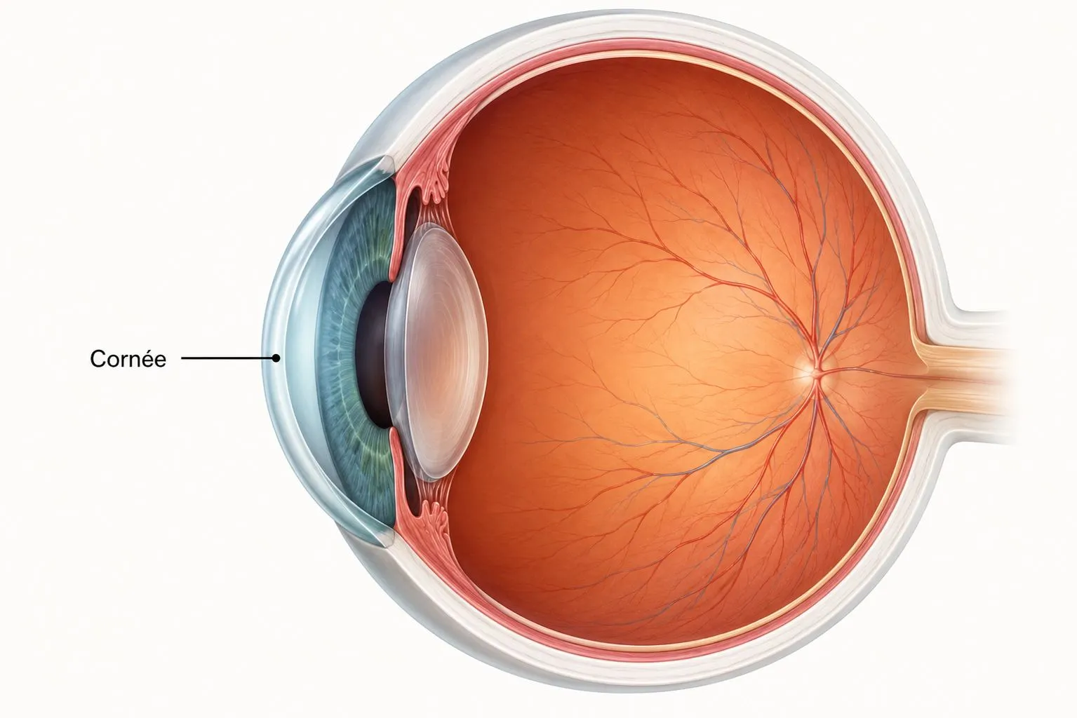
The cornea is the transparent lens at the front of the eye; normally smooth and spherical, it provides most of the focusing of images. In keratoconus, it gradually thins and becomes deformed, losing its roundness and taking on the shape of an irregular cone (from the Greek kerato, cornea, and konos, cone).
This deformation creates a progressive irregular astigmatism: images become blurred and distorted, and are poorly corrected by simple glasses. Keratoconus most often affects both eyes, asymmetrically. It generally begins in adolescence or in young adults and progresses mainly before the age of 40, before stabilising.
Symptoms that should alert you
Keratoconus begins discreetly. Certain signs, especially in a teenager or young adult, should prompt a consultation:
The role of eye rubbing
We now know that repeated eye rubbing plays a major role: the mechanical stress it causes weakens the cornea and encourages it to bulge. This is why the first step, at any stage, is to stop rubbing your eyes, a simple action that helps slow progression.
Other factors are associated: an allergic background (atopy, which precisely leads people to rub their eyes), a family history of keratoconus, and more rarely certain general diseases of the connective tissue. Treating an eye allergy is therefore an integral part of management.
Diagnosis
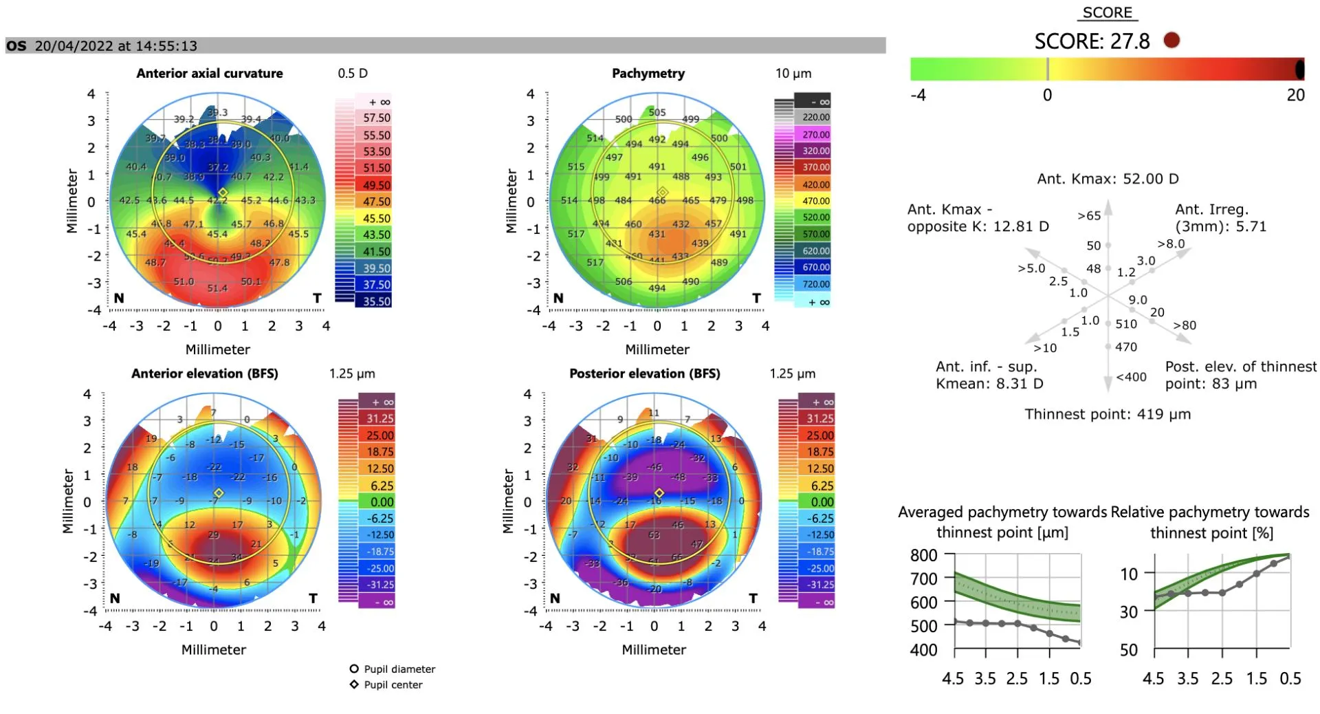
Early keratoconus can go unnoticed. Its diagnosis and monitoring rely on precise tests, carried out at the practice:
Treatments
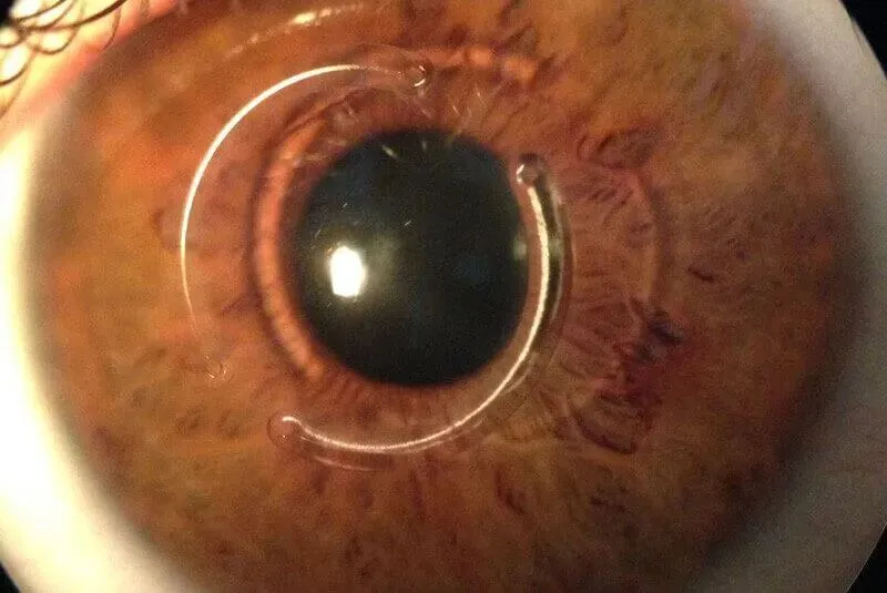
Management is stepwise and adapted to the stage: first correct vision, and above all stabilise the cornea when the keratoconus is progressing.
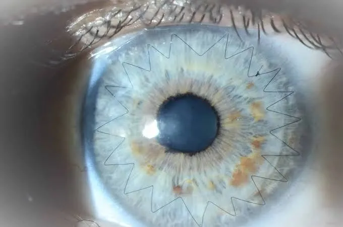
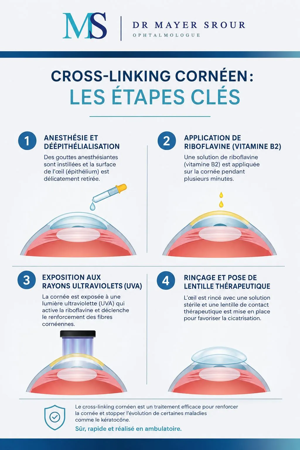
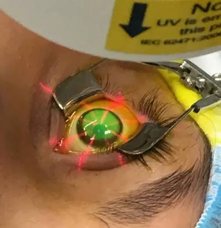
Detected early, keratoconus stabilises in the great majority of cases. Everything depends on early diagnosis, stopping eye rubbing and timely cross-linking; a transplant concerns only a minority of patients.
Keratoconus in Noisy-le-Grand
The Grand Paris Ophtalmologie practice provides screening, topographic monitoring and management of keratoconus on site, for patients from Noisy-le-Grand and eastern Paris (93, 94, 77): Bry-sur-Marne, Villiers-sur-Marne, Champs-sur-Marne, Chelles, Torcy and Marne-la-Vallée, as well as Le Perreux-sur-Marne, Nogent-sur-Marne, Fontenay-sous-Bois, Vincennes, Saint-Maur-des-Fossés, Créteil, Montreuil and Rosny-sous-Bois. Follow-up is provided by Dr Yaïr Chetrit or Dr Mayer Srour, using the practice's corneal topography.
Keratoconus: your questions
Answers from Dr Yaïr Chetrit and Dr Mayer Srour, ophthalmologists in Noisy-le-Grand.
Does keratoconus cause blindness?
No. Even when advanced, keratoconus does not cause blindness: central vision is affected but peripheral vision is preserved. Detected early and stabilised, it allows a good quality of life.
Can I have laser surgery (LASIK)?
No: keratoconus is a contraindication to laser vision correction, which would thin the cornea further. This is why topography is routinely performed before any refractive surgery.
Is keratoconus hereditary?
There is a family tendency: about one patient in five has an affected relative. If a member of your family has keratoconus, screening by topography is advised, especially in adolescence.
Is cross-linking painful?
The session is carried out under drop anaesthesia and is not painful. Discomfort and a gritty sensation are common for the following 2 to 3 days, while the surface heals under a bandage lens.
How do I know if it is progressing?
By comparing topographies from one visit to the next. If the cone becomes more pronounced, cross-linking is offered to stabilise the cornea before vision deteriorates.
Why shouldn't I rub my eyes?
Because repeated rubbing puts mechanical stress on the cornea, which deforms it and worsens keratoconus. Stopping eye rubbing, and treating any allergy, is an essential step.
At what age does it appear?
Most often in adolescence or young adulthood. It progresses mainly until about 40 years of age, then tends to stabilise, hence the importance of early screening and follow-up.
Can wearing lenses cure keratoconus?
No: rigid lenses correct vision very well by smoothing out irregularities, but they do not stop progression. Only cross-linking stabilises the cornea when the disease is progressing.
Related reading
Reduced vision or doubts about keratoconus?
Screening and follow-up at the Noisy-le-Grand practice, with corneal topography on site.
The Grand Paris Ophtalmologie practice in Noisy-le-Grand provides consultations, screening and surgery for patients from neighbouring towns: