

OCT: the scan of your retina
OCT produces cross-sections of your retina accurate to the micron, like a scanner — with no contact and no X-rays. It is the key examination for monitoring AMD, diabetes and glaucoma.
OCT: the essentials in 30 seconds
- OCT (optical coherence tomography) is a retinal scan using infrared light: no contact, no injection, no X-rays, a few seconds per eye.
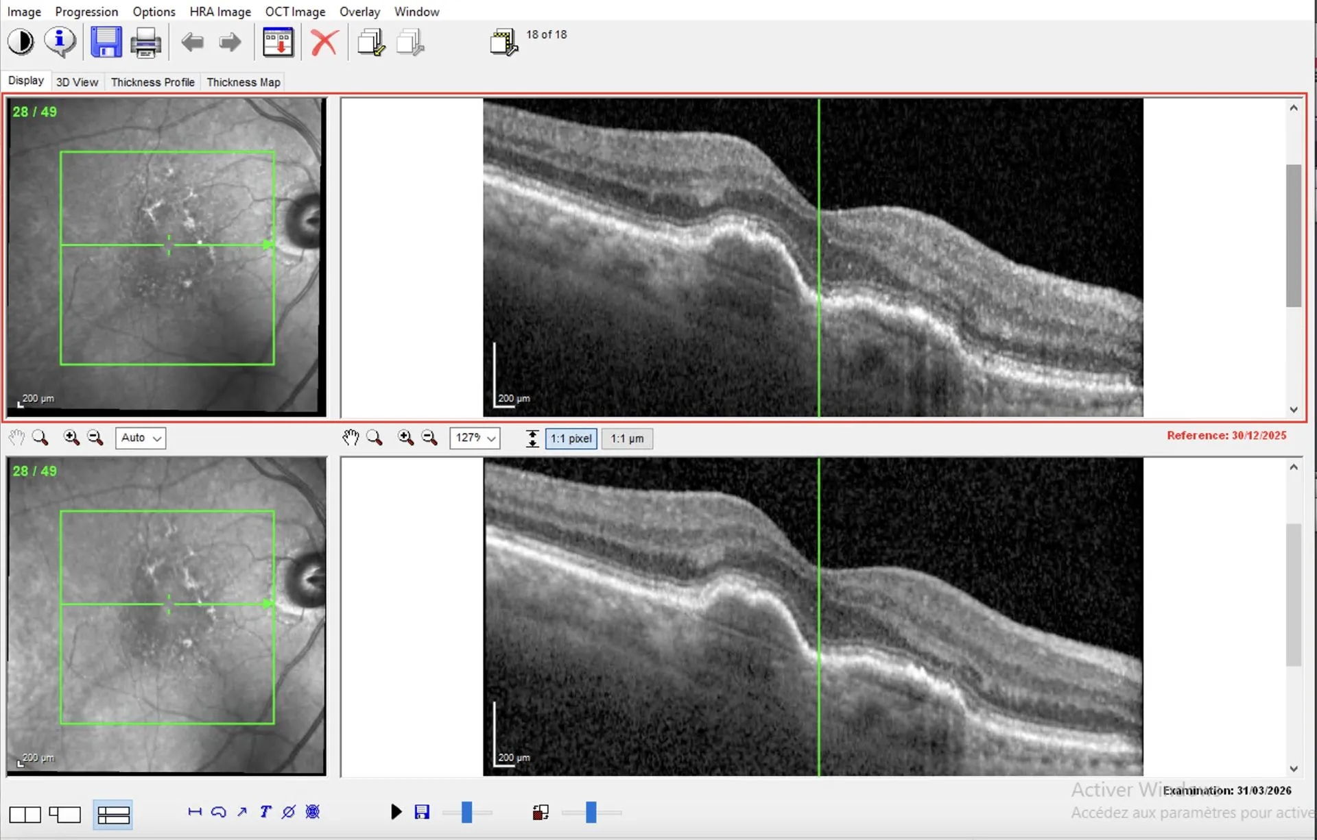
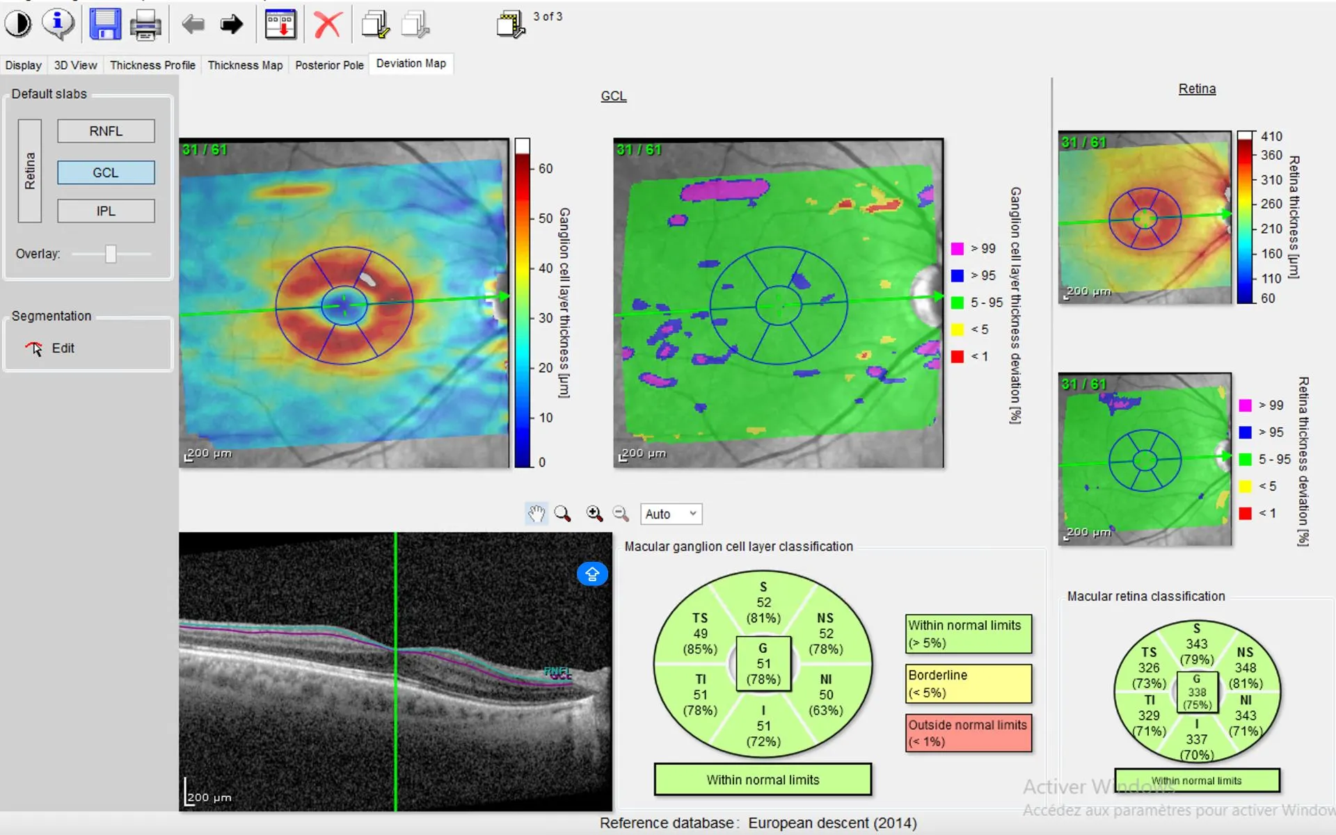
- Macular OCT: it analyses the macula, the centre of the retina. Essential for AMD, diabetic oedema, macular membranes and holes.
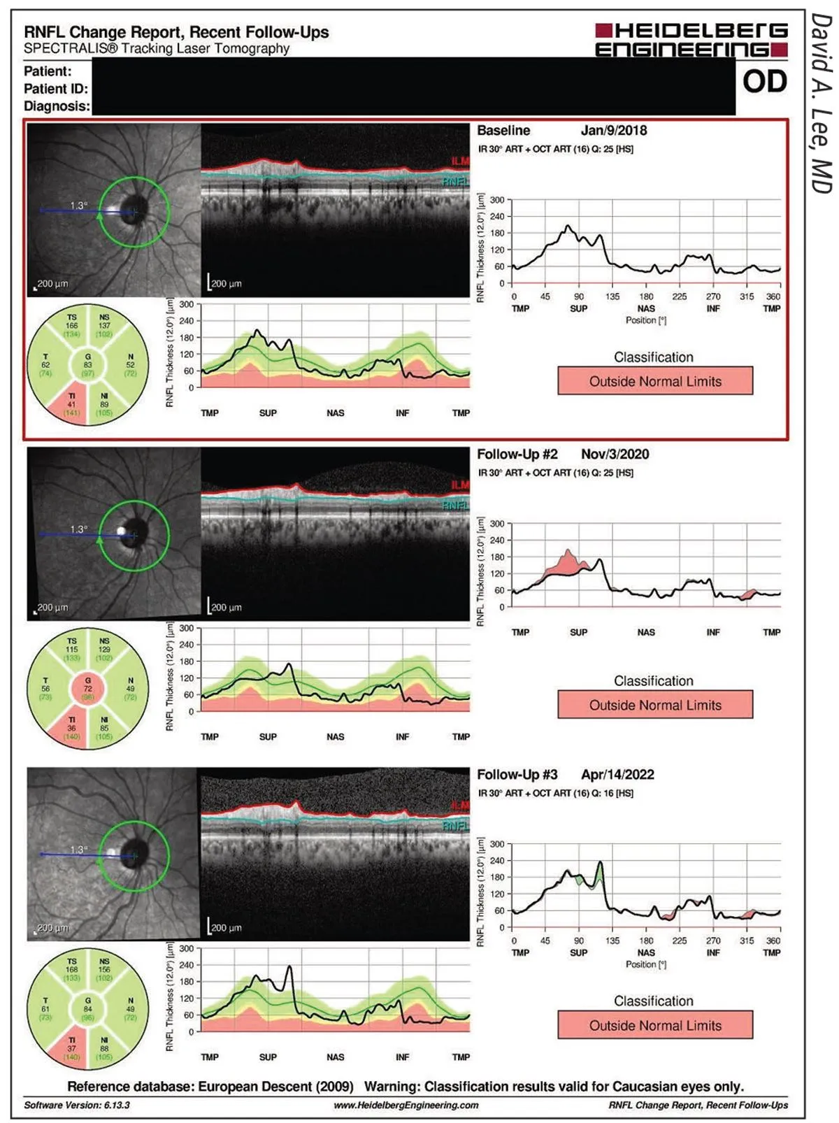
- RNFL OCT (optic nerve): it measures the thickness of the optic nerve fibres. It is the key examination for screening and monitoring glaucoma, often well before the visual field.
- A normal macular OCT shows a regular profile with a central depression (the fovea), clear layers and a thickness within the device’s normal range.
What is OCT?
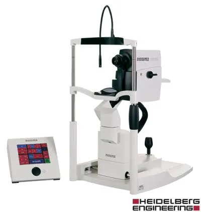
OCT (optical coherence tomography) is an imaging examination that produces cross-sections of the retina accurate to the micron — like a scanner, but using a simple beam of light, with no contact, no injection and no X-rays. In a few seconds, it reveals the different layers of the retina, which are invisible on a fundus examination.
The practice also has OCT angiography, which shows the vessels of the retina without injecting dye — a valuable complement to conventional angiography.
Macular OCT or optic nerve OCT?
The same device performs two complementary analyses, depending on what your doctor wishes to explore:
What happens during the examination?
A normal OCT: what does the result look like?
Normal macular OCT: the profile is regular, with a small depression at the centre (the fovea), clear retinal layers and a thickness within the device’s normal values.
Normal RNFL OCT: the thickness of the optic nerve fibres is within normal values for age, and the two eyes are close to one another. The report classifies each sector by colour: green = normal, yellow = borderline, red = outside normal limits.
A result is always read together with the clinical examination: only the ophthalmologist can interpret it.
OCT in Noisy-le-Grand
The Grand Paris Ophtalmologie practice is equipped with a latest-generation OCT with an angiography module, for patients from Noisy-le-Grand and eastern Paris (93, 94, 77): Bry-sur-Marne, Villiers-sur-Marne, Champs-sur-Marne, Chelles, Torcy and Marne-la-Vallée, as well as Le Perreux-sur-Marne, Nogent-sur-Marne, Fontenay-sous-Bois, Vincennes, Saint-Maur-des-Fossés, Créteil, Montreuil and Rosny-sous-Bois. The examination is performed and interpreted on site by Dr Yaïr Chetrit or Dr Mayer Srour — from the first consultation if needed.
OCT: your questions
Answers from Dr Yaïr Chetrit and Dr Mayer Srour, ophthalmologists in Noisy-le-Grand.
Is the test painful?
No — OCT is completely painless and contact-free: you simply look at a light target for a few seconds. No injection, no X-rays.
Macular OCT or RNFL optic nerve OCT: what is the difference?
Macular OCT explores the centre of the retina (AMD, diabetic oedema); optic nerve OCT measures the nerve fibres (glaucoma). Same device, two analyses.
Is an injection or dilation needed?
No injection — that is the whole point of OCT angiography. Dilation is not routine: it is only performed for a more complete examination of the retina.
How often should an OCT be done?
Depending on the condition: very regularly for wet AMD or diabetic oedema treated with injections; 1 to 2 times a year to monitor glaucoma, comparing the examinations.
Are OCT and angiography the same thing?
No: conventional angiography injects a dye to film the circulation. OCT angiography shows the vessels without injection — the two are complementary.
Can I drive afterwards?
Yes if the examination was done without dilation. If dilation was performed, bring someone with you: the glare lasts a few hours.
What is a normal macular OCT?
On a normal macular OCT, the layers of the retina are regular and well aligned, the fovea forms a small central depression, there is no fluid (oedema), no detachment and no membrane on the surface, and the central thickness is within the reference values (around 250 microns depending on the device). Only the ophthalmologist interprets the result in light of your symptoms.
What is the RNFL in ophthalmology?
The RNFL (Retinal Nerve Fibre Layer) is the layer of nerve fibres in the retina that form the optic nerve. RNFL OCT measures its thickness around the optic disc: thinning, especially if asymmetric or localised, is an early sign of glaucoma. Compared from one examination to the next, it shows whether the disease is progressing.
Is OCT a scan? Do I need a prescription?
OCT is often called an “optic nerve scan” or “retinal scan”, but it uses no X-rays, only light. It is performed directly at the practice, during the consultation, with no prescription or special preparation.
Related reading
Have you been prescribed an OCT?
The examination is carried out at the Noisy-le-Grand practice and interpreted immediately.
The Grand Paris Ophtalmologie practice in Noisy-le-Grand provides consultations, screening and surgery for patients from neighbouring towns: