
Corneal topography
Topography produces a precise map of the shape and thickness of the cornea. A key examination before refractive surgery, to detect keratoconus and to fit custom contact lenses.
What is corneal topography?
The cornea is the transparent lens at the front of the eye; it provides most of the focusing of images. Corneal topography produces a precise map of it: its curvature, its regularity and its thickness, point by point.
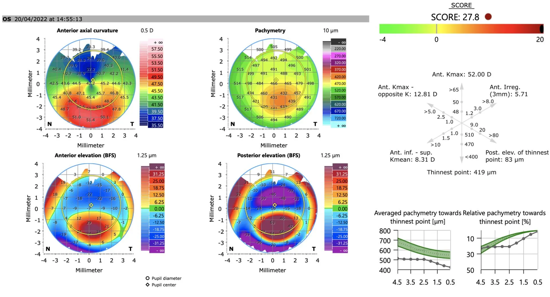
The result takes the form of a colour map — red for steeper areas, blue for flatter ones. It reveals the slightest irregularity, often invisible on a simple examination, such as early keratoconus.
What can it detect?
A single measurement is not enough: it is the complete map that matters. Topography reveals:
Topography or tomography?
The two terms are close, but do not describe exactly the same thing. Topography studies the surfaces of the cornea — the curvature of its front and back faces. Tomography goes further: it reconstructs the cornea through its whole thickness and across its whole surface, providing a complete three-dimensional analysis.
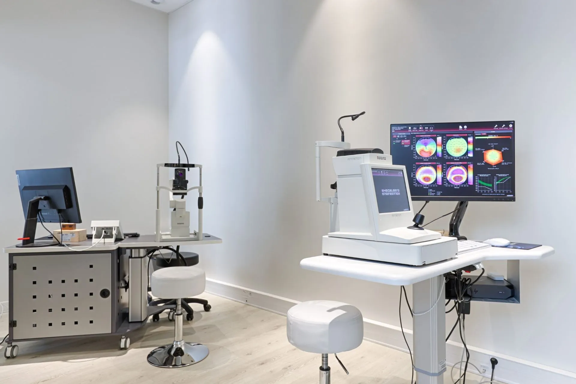
The practice's modern devices combine both — depending on the technology, a slit of light, a beam or concentric rings scan the cornea — to deliver a complete set of curvature, elevation and thickness maps.
When is it useful?
What happens during the examination?
It could not be simpler: seated, with your chin resting on the device, you are asked to blink several times to even out the tear film, then to look at a target without moving for a few seconds while the device scans the cornea. No contact, no pain and no dilation — the examination takes only a few minutes. The maps are interpreted immediately by your ophthalmologist, then kept so they can be compared from one visit to the next.
Corneal topography in Noisy-le-Grand
The Grand Paris Ophtalmologie practice carries out corneal topography on site, for patients from Noisy-le-Grand and eastern Paris (93, 94, 77): Bry-sur-Marne, Villiers-sur-Marne, Champs-sur-Marne, Chelles, Torcy and Marne-la-Vallée, as well as Le Perreux-sur-Marne, Nogent-sur-Marne, Fontenay-sous-Bois, Vincennes, Saint-Maur-des-Fossés, Créteil, Montreuil and Rosny-sous-Bois. The examination is carried out and interpreted by Dr Yaïr Chetrit or Dr Mayer Srour, in particular as part of the refractive surgery assessment.
Corneal topography: your questions
Answers from Dr Yaïr Chetrit and Dr Mayer Srour, ophthalmologists in Noisy-le-Grand.
Is it painful?
Not at all: the examination is painless and most often contactless. You simply look at a target for a few seconds, without dilation.
Why before refractive surgery?
Because it checks that the cornea is regular and thick enough and rules out keratoconus, the main contraindication to laser treatment. It is a safety examination in the pre-operative assessment.
Can it detect keratoconus?
Yes, it is the reference examination for detecting it even at an early stage and following it over time by comparing successive maps.
Are topography and pachymetry the same thing?
They are two complementary measurements, often obtained with the same device: topography maps the shape of the cornea, while pachymetry measures its thickness.
Related reading
Have you been prescribed corneal topography?
The examination is carried out at the Noisy-le-Grand practice and interpreted immediately.
The Grand Paris Ophtalmologie practice in Noisy-le-Grand provides consultations, screening and surgery for patients from neighbouring towns: