

The Eidon: the retina photographed in true colour, right out to the periphery
The Eidon takes our ultra-widefield retinal photographs: images of the retina in true colour, from the centre to the periphery, most often without dilation — the ideal screening tool.
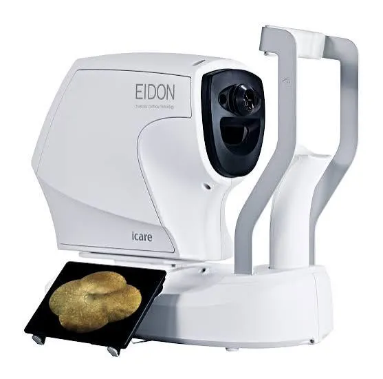
What is the Eidon, and what is it used for?
The Eidon is a confocal retinal camera: it photographs the back of your eye — retina, macula, optic nerve, vessels — in high resolution. Where a standard retinal photograph shows only a central area of about 45°, the Eidon captures a very wide panorama, extended to the periphery by automatically stitching images together (mosaic).
Each image is archived: from one visit to the next, the doctor compares the images side by side and immediately spots what has changed — a haemorrhage, a new naevus, a peripheral lesion.
Why it is a benchmark in its category
Its confocal technology retains only the light reflected from the plane of the retina: the images are sharp, high-contrast and in true colour — faithful to what the doctor's own eye would see — even through a small pupil or an early cataract. The flash is gentle and not dazzling.
This is what makes it the ideal screening tool: most examinations are done without dilation, so no blurred vision afterwards — you can drive home. And the ultra-wide field reveals peripheral lesions (tears, areas of ischaemia in diabetes) that are invisible on a standard image.
When the practice uses it
What it means for you
The Eidon: your questions
Answers from Dr Yaïr Chetrit and Dr Mayer Srour, ophthalmologists in Noisy-le-Grand.
Does the pupil need to be dilated for the examination?
Most often no: the Eidon photographs through a normal pupil. Dilation remains necessary in certain cases (a very peripheral examination) — the doctor will let you know.
Is the examination dazzling?
Very little: the confocal flash is gentle, much more comfortable than a conventional retinal photograph. The glare fades within a few seconds.
Does it replace the doctor's fundus examination?
It complements it: the photo documents and archives, the doctor interprets — and keeps the clinical examination (with dilation if needed) for situations that require it.
Related reading
Need a screening fundus examination?
The Eidon is installed at the Noisy-le-Grand practice — the examination takes a few seconds per eye, most often without dilation.
The Grand Paris Ophtalmologie practice in Noisy-le-Grand provides consultations, screening and surgery for patients from neighbouring towns: