

Retinal surgery
Some diseases of the retina and the macula require surgery. Retinal detachment is an emergency; epiretinal membrane and macular hole distort central vision and are corrected by microsurgery. At the Noisy-le-Grand practice, Dr Mayer Srour and Dr Yaïr Chetrit provide diagnosis, imaging and follow-up, in liaison with retinal surgery centres.
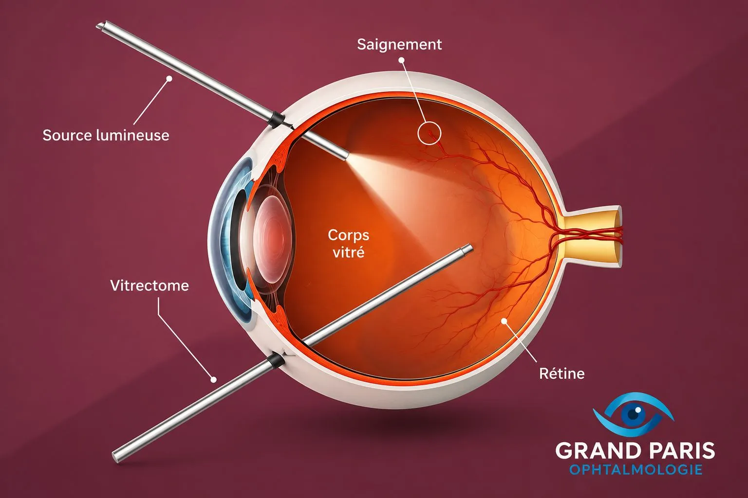
Retinal surgery: the essentials in 30 seconds
- Retinal detachment is a surgical emergency: the earlier the operation, the better the recovery.
- Warning signs: flashes of light, a shower of floaters, a black veil that spreads.
- Surgery (vitrectomy) also treats epiretinal membranes and macular holes, which cause visual distortion.
- Surgery performed by Dr Srour and Dr Chetrit; full assessment at the practice (OCT, ultrasound) before any decision.
Retinal detachment — an emergency
The retina, the thin membrane lining the back of the eye, can lift away from the wall — most often from a peripheral tear through which fluid passes under the retina. Without treatment, the detachment spreads and vision may be permanently lost. It is a surgical emergency.
Warning signs: the sudden appearance of flashes of light, a shower of floaters, then a black veil or curtain that hides part of your vision. If you notice these signs, seek urgent advice.
Treatment is surgical and adapted to the type of detachment: most often a vitrectomy (see below), sometimes supplemented by gas or silicone oil to hold the retina in place, or by a scleral buckle (a small sponge placed on the wall of the eye). A simple tear, detected early, is treated with laser and may prevent detachment. High myopia carries a higher risk.
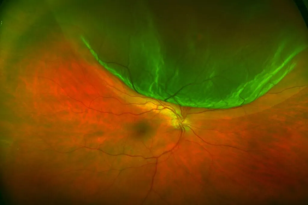
Understanding retinal detachment: the role of the vitreous, the tear, the warning signs and why you must be seen urgently.
Find all of Dr Srour’s videos on his YouTube channel: Subscribe to the channel
Epiretinal membrane
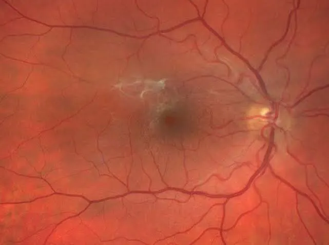
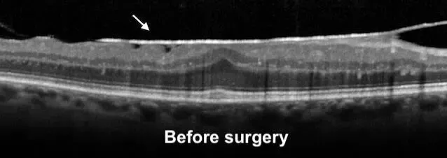
A thin film forms on the surface of the macula (the centre of vision) and contracts, like a layer of cellophane puckering the retina. Common after 60, often with no specific cause, it causes distorted central vision (straight lines look wavy) and gradually blurred vision.
The diagnosis is confirmed by OCT, which measures the thickness of the macula and the effect of the membrane. As long as symptoms are mild, simple monitoring is enough.
When symptoms become significant, a vitrectomy with peeling gently removes the membrane from the surface of the retina. Vision gradually improves over the following months (recovery depends on how long the membrane has been present).
Macular hole
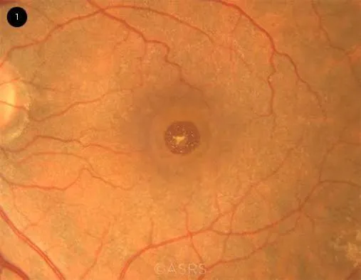
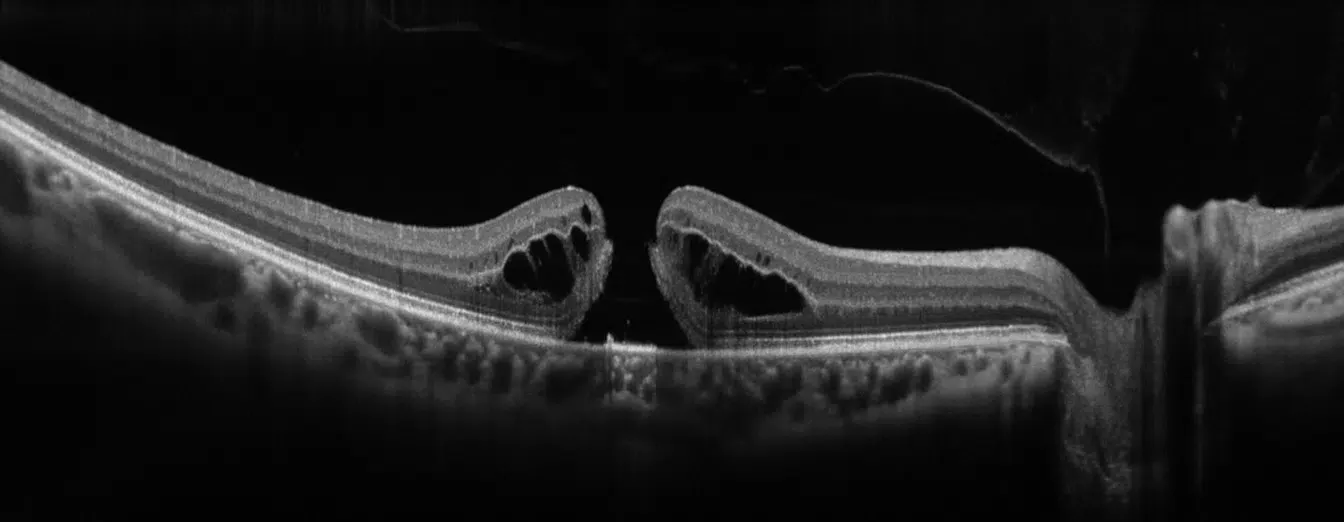
A macular hole is a small opening that forms at the exact centre of the macula, often linked to traction from the vitreous as we age. It causes a central blind spot, distortion of images and a loss of fine vision (reading, faces).
Here again, OCT makes the diagnosis precisely and measures the size of the hole, which guides the decision.
Vitrectomy with peeling and gas tamponade makes it possible to close the hole in the great majority of cases. A face-down position is sometimes requested for a few days after the operation, while the gas bubble does its work.
Vitrectomy in practice
Vitrectomy is the reference microsurgery of the retina. Through tiny incisions (less than a millimetre), the surgeon removes the vitreous gel that fills the eye in order to reach the retina: removing a membrane, closing a hole, reattaching the retina.
At the Noisy-le-Grand practice, Dr Srour and Dr Chetrit make the diagnosis (OCT, imaging), schedule the operation in liaison with retinal surgery centres, and provide follow-up.
Retinal surgery: the right reflexes
Flashes, a shower of floaters, then a black veil: signs of retinal detachment. Be seen the same day.
Wavy lines, a central blind spot: a membrane or a macular hole can be detected in a few minutes with OCT.
Detected early, a simple tear is treated with laser and often avoids detachment surgery.
After a gas tamponade, follow the requested positioning and do not fly while gas remains.
Official factsheets from the Société Française d'Ophtalmologie
Written by experts of the SFO, they detail the procedure, benefits and risks of retinal surgery. To be read before your operation.
Retinal surgery in Noisy-le-Grand
The Grand Paris Ophtalmologie practice provides diagnosis and follow-up of surgical retinal diseases (retinal detachment, epiretinal membrane, macular hole) for patients from Noisy-le-Grand and eastern Paris (93, 94, 77): Bry-sur-Marne, Villiers-sur-Marne, Champs-sur-Marne, Neuilly-sur-Marne, Gournay-sur-Marne, Noisiel, Torcy, Chelles, Gagny, Marne-la-Vallée, but also Le Perreux-sur-Marne, Nogent-sur-Marne, Vincennes, Saint-Maur-des-Fossés, Créteil, Rosny-sous-Bois and Montreuil.
Dr Mayer Srour, trained at the CHI de Créteil, and Dr Yaïr Chetrit provide diagnosis and imaging at the practice and schedule the operation in liaison with retinal surgery centres. In an emergency, contact us: flashes, floaters: seek urgent advice.
Retinal surgery: your questions
Answers from Dr Mayer Srour and Dr Yaïr Chetrit, ophthalmologists in Noisy-le-Grand.
What are the signs of retinal detachment?
The sudden appearance of flashes of light, a shower of floaters, then a black veil or curtain in the field of vision. It is an emergency: be seen the same day.
Can retinal detachment be treated?
Yes, with surgery (most often a vitrectomy), all the better when it is carried out quickly. The result depends greatly on how early it is done and on whether the centre of vision is affected.
What is an epiretinal membrane?
A thin film that puckers the macula and distorts central vision. When symptoms are significant, it is removed by peeling (vitrectomy).
Can a macular hole close?
Yes: surgery (vitrectomy, peeling and gas) closes the hole in the great majority of cases, with gradual improvement in vision.
What is a vitrectomy?
The reference surgery of the retina: through tiny incisions, the vitreous gel is removed in order to work on the retina (peeling, closing a hole, reattachment).
Do I need to stay in a particular position after the operation?
After a gas tamponade, a head position (often face down) may be requested for a few days. You cannot travel by air until the gas has been absorbed.
Is the operation painful?
No. It is performed under anaesthesia (local or general) and is not painful. A few days of discomfort and eye drops follow, with close check-ups.
Where is the surgery performed?
Diagnosis and follow-up (OCT, imaging) are provided at the Noisy-le-Grand practice; the operation is scheduled in the operating theatre, in liaison with retinal surgery centres.
Related reading
Flashes, floaters, black veil? Don't wait.
Diagnosis of surgical retinal diseases at the Noisy-le-Grand practice — retinal detachment is an emergency.
The Grand Paris Ophtalmologie practice in Noisy-le-Grand provides consultations, screening and surgery for patients from neighbouring towns: