

Posterior vitreous detachment
With age, the gel (the vitreous) that fills the eye gradually separates from the retina. It is a little as if the jelly inside your eye decided to move a little away from the retina: it often happens with age and is generally not serious if there is no retinal tear. At the Noisy-le-Grand practice, Dr Mayer Srour and Dr Yaïr Chetrit check the retina and rule out a tear.
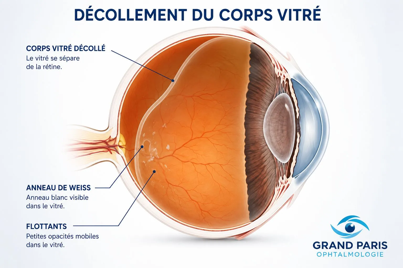
Vitreous detachment: the essentials in 30 seconds
- How long does a vitreous detachment last? Floaters and flashes most often fade within a few weeks to a few months, but some floaters can persist for longer. A fundus check-up at 4–6 weeks is systematically recommended.
- PVD is a normal ageing change of the vitreous gel, common between 50 and 70.
- Harmless in the great majority of cases — but a dilated fundus examination within the following days is essential to rule out a retinal tear.
- Immediate emergency if: a shower of black dots, repeated flashes, a dark veil in the field of vision.
- Floaters fade with time; no treatment is needed in the simple form.
What is posterior vitreous detachment?
The inside of the eye is filled with a transparent gel, the vitreous, attached to the retina. With age, this gel shrinks and eventually detaches from the surface of the retina: this is posterior vitreous detachment (PVD). In other words, the jelly filling your eye moves a little away from the retina.
It is a natural and very common process after the age of 55–60, affecting one eye and then often the other, a few months or years apart. In the great majority of cases it is harmless and needs no treatment — provided there is no retinal tear. This is precisely what the fundus examination checks.
Floaters & flashes of light
Vitreous detachment shows itself through two typical symptoms, often appearing fairly suddenly:


How long does a vitreous detachment last? The duration varies greatly from person to person. For some, floaters and flashes ease within a few weeks or months; for others, they persist for longer. The brain most often learns to ignore them and they become less bothersome. We systematically recommend a check-up at the practice at 4–6 weeks.
The 3 signs that mean you must be seen again urgently
In a minority of cases, a vitreous detachment can cause a retinal tear — the gateway to a retinal detachment, which is an emergency. These three signs should prompt you to be seen the same day:
In that case, call 01 85 10 57 00 — emergencies are seen the same day.
Diagnosis, in a single visit
The essential test is the dilated fundus examination: it shows the vitreous detachment and, above all, allows us to check the whole periphery of the retina for a tear. If needed, it is completed using the practice's on-site equipment:
If no tear is found, the PVD is harmless: we reassure you and schedule a check-up. If a tear is found, it is treated the same day with laser.
Course & monitoring
An isolated PVD is not treated. There is no medicine or surgery to make the floaters of a simple vitreous detachment disappear: they fade with time and the brain learns to ignore them. What to do is simple:
Simple monitoring: systematic fundus check at 4–6 weeks, then a consultation if any new symptom appears. No treatment needed.
A barrier laser (photocoagulation) is performed at the practice around the tear to “weld” the retina and prevent retinal detachment.
Discover medical laser treatmentVitreous detachment: good habits
Recent floaters or flashes call for a dilated fundus examination, to rule out a tear.
A sudden increase in floaters, new flashes or a black veil: be seen the same day.
Without a tear, there is no treatment: floaters fade and the brain learns to ignore them.
We systematically recommend a fundus check-up at 4–6 weeks, once the detachment has run its course.
The official information sheet from the Société Française d'Ophtalmologie
If a tear is treated, retinal laser treatment is described in the official SFO information sheet — worth reading before your session.
Floaters & flashes: seeing a doctor in Noisy-le-Grand
The Grand Paris Ophtalmologie practice promptly sees patients troubled by recent floaters or flashes of light, in Noisy-le-Grand and throughout eastern Paris (93, 94, 77): Bry-sur-Marne, Villiers-sur-Marne, Champs-sur-Marne, Chelles, Torcy, Marne-la-Vallée, but also Le Perreux-sur-Marne, Nogent-sur-Marne, Vincennes, Saint-Maur-des-Fossés, Créteil, Montreuil and Rosny-sous-Bois — fundus examination, imaging and laser on site.
Dr Mayer Srour, trained at the CHI de Créteil, and Dr Yaïr Chetrit examine the periphery of the retina and rule out a tear at the first visit. In an emergency, contact us: flashes, floaters: seek urgent care.
Vitreous detachment: your questions
Answers from Dr Mayer Srour and Dr Yaïr Chetrit, ophthalmologists in Noisy-le-Grand.
Is vitreous detachment serious?
No, in the vast majority of cases. It is a natural, age-related process with no consequences, as long as there is no retinal tear. This is precisely what the fundus examination checks.
Do floaters go away?
They most often become less bothersome with time: they fade, drift out of the line of sight and the brain learns to ignore them. They do not always disappear completely, but significant, persistent discomfort should be reassessed.
What signs should alert me?
Three signs mean you should be seen the same day: a sudden increase in floaters, the appearance of new flashes of light, or a black veil in the field of vision — they may point to a retinal tear.
How long do the symptoms last?
The duration varies greatly: a few weeks to a few months for most people, sometimes longer. A check-up at 4–6 weeks is systematically recommended to make sure everything is stable.
Is treatment needed?
An isolated PVD is not treated: there are no eye drops or surgery for the floaters of a simple detachment. However, an associated tear is treated the same day with laser.
Can vitreous detachment affect both eyes?
Yes: it occurs in each eye, often a few months or years apart. After the first eye, any new symptom in the other eye warrants the same fundus examination.
Who is most affected?
Mainly after 55–60. The risk comes earlier in cases of high myopia, after cataract surgery or an injury.
Where can I be seen for floaters near Noisy-le-Grand?
At the Grand Paris Ophtalmologie practice, 67 rue de Malnoue in Noisy-le-Grand (93160). Dilated fundus examination, OCT and laser if necessary are carried out on site. Telephone: 01 85 10 57 00.
Related reading
Recent floaters or flashes? Be seen quickly.
Fundus examination, imaging and laser at the Noisy-le-Grand practice — emergencies seen the same day.
The Grand Paris Ophtalmologie practice in Noisy-le-Grand provides consultations, screening and surgery for patients from neighbouring towns: