

Diabetes and the eyes: diabetic retinopathy
Everyone with diabetes should have their fundus checked once a year, even with no visual symptoms: diabetic retinopathy progresses silently and is easier to treat the earlier it is detected. The examination (retinal photography, OCT) is painless and carried out on site in a single consultation.
Diabetes damages the eyes silently: one fundus examination a year protects your vision. At the Noisy-le-Grand practice, Dr Mayer Srour — — and Dr Yaïr Chetrit provide screening, treatment and follow-up, in coordination with your diabetologist.

Diabetic retinopathy: the essentials in 30 seconds
- One of the leading causes of visual impairment in adults under 65 — and for a long time without any symptoms.
- One fundus examination a year for everyone with diabetes, even with perfect vision.
- Keeping blood sugar and blood pressure under control remains the first treatment.
- If necessary: laser or injections — all the more effective when the disease is detected early.
What is diabetic retinopathy?
Chronically excessive blood sugar weakens the walls of the small vessels that nourish the retina. Over time, these vessels can leak, become blocked or grow abnormally: this is diabetic retinopathy, which affects both type 1 and type 2 diabetes.
Its danger: it progresses for years without any symptoms. By the time vision drops, the disease is already advanced. It is the leading cause of blindness before the age of 50 in industrialised countries — and yet, when detected and treated in time, more than 90% of severe vision loss is avoidable.
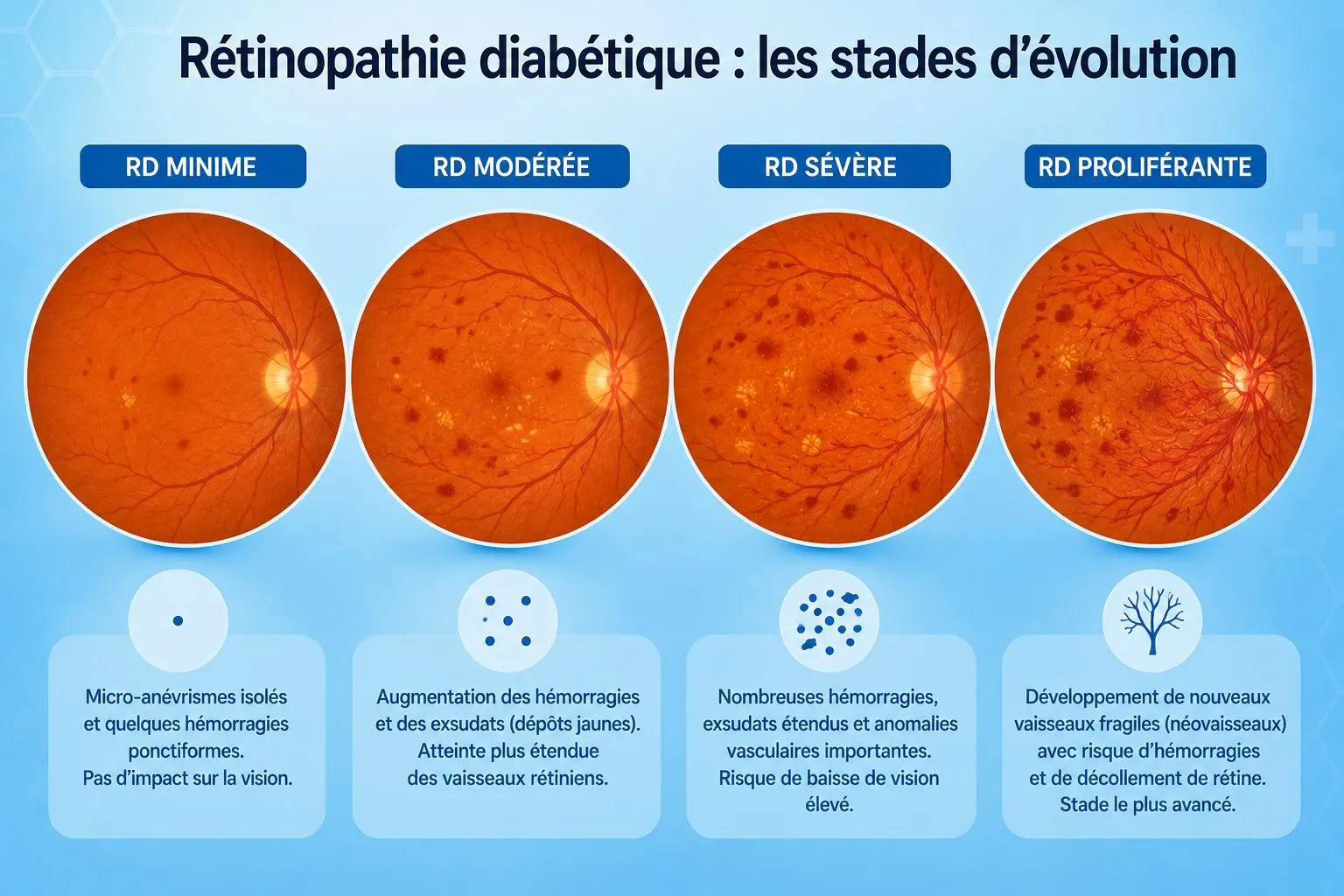
The stages of the disease
Retinopathy progresses through stages of increasing severity — each stage determines the monitoring schedule and the treatment:
Screening: one fundus examination a year
When retinopathy is already known, the schedule is tightened according to the stage: every 6 months, 3–4 months, up to monthly during active treatment.
At the practice, the assessment is carried out in a single visit on the technical platform: ultra-widefield retinal photography (more than 80% of the retina in one image), macular OCT, OCT-angiography without injection and, if needed, fluorescein angiography.
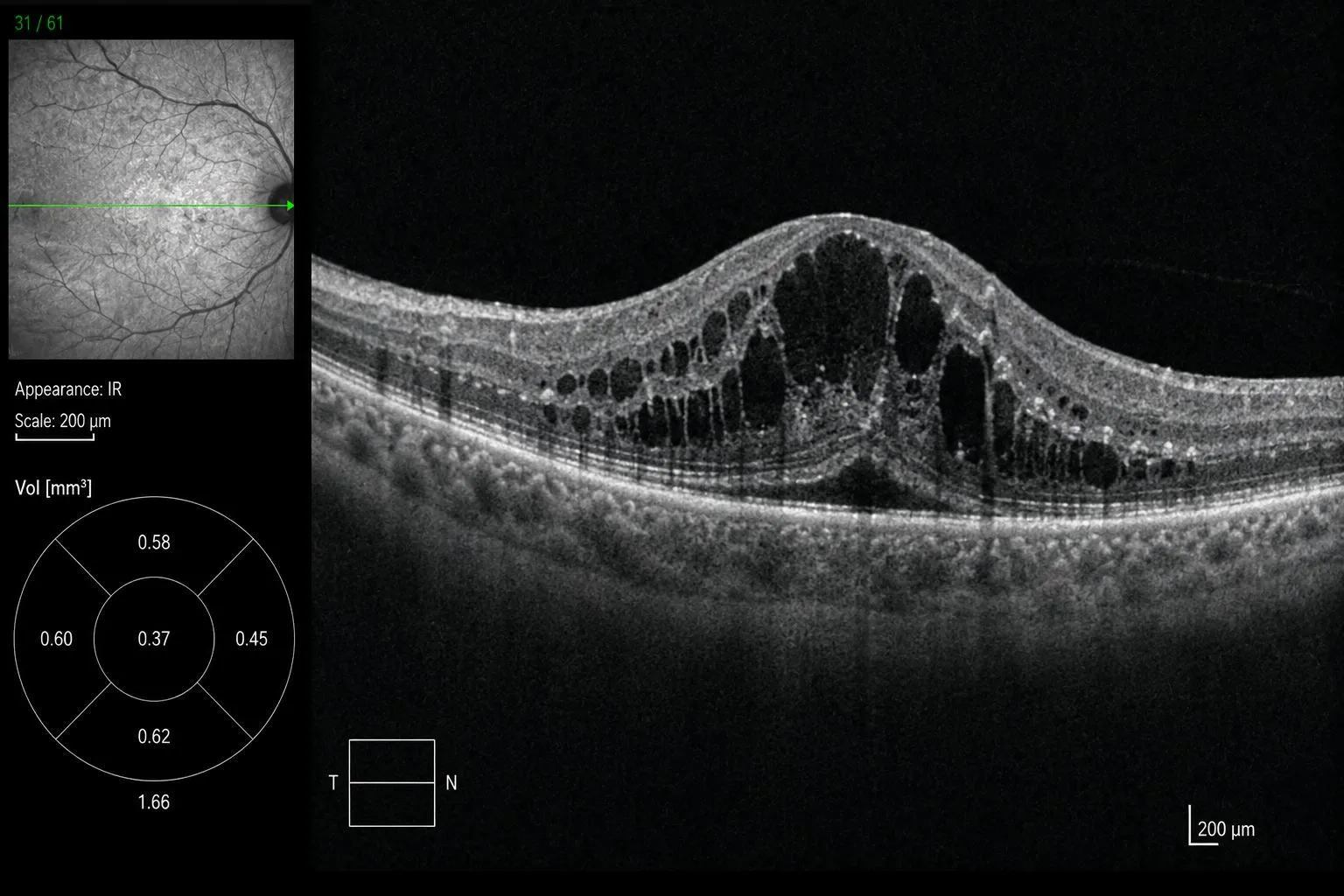
Diabetic macular oedema
When weakened vessels leak at the centre of the retina, fluid builds up in the macula: this is diabetic macular oedema, the leading cause of vision loss in people with diabetes.
It is detected by macular OCT — painless, contactless, a few minutes — and effectively treated with intravitreal injections carried out at the practice, in the dedicated room.
Treatments
The foundation: controlling the diabetes. Blood sugar (HbA1c), blood pressure and cholesterol are the first treatments for the retina — in close coordination with your diabetologist and your GP (médecin traitant).
Depending on the stage, two key treatments are carried out directly at the practice — surgical forms are operated on in a partner clinic, with follow-up on site:
Photocoagulation with Argon laser: a session at the practice, seated at the slit lamp, under numbing eye drops with a contact lens. It treats ischaemic areas, prevents complications of the proliferative form and seals tears.
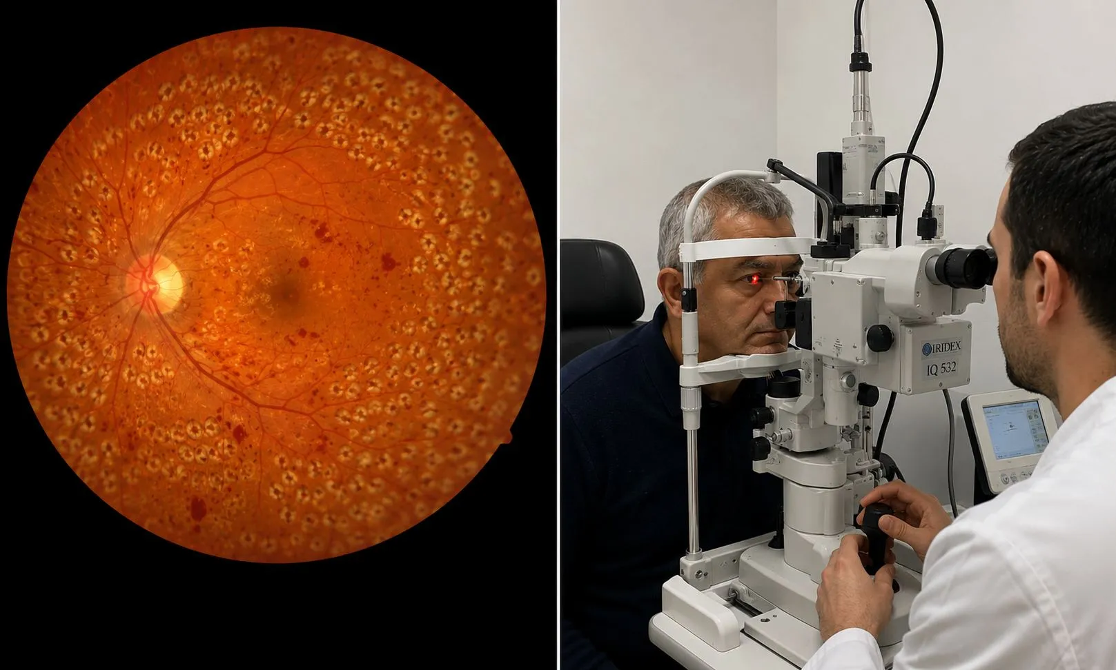 Discover the practice’s laser equipment
Anti-VEGF or corticosteroids for macular oedema: a few seconds, under numbing eye drops, in the practice's dedicated room with strict aseptic conditions — no hospital stay. Schedule tailored to the OCT check-ups.
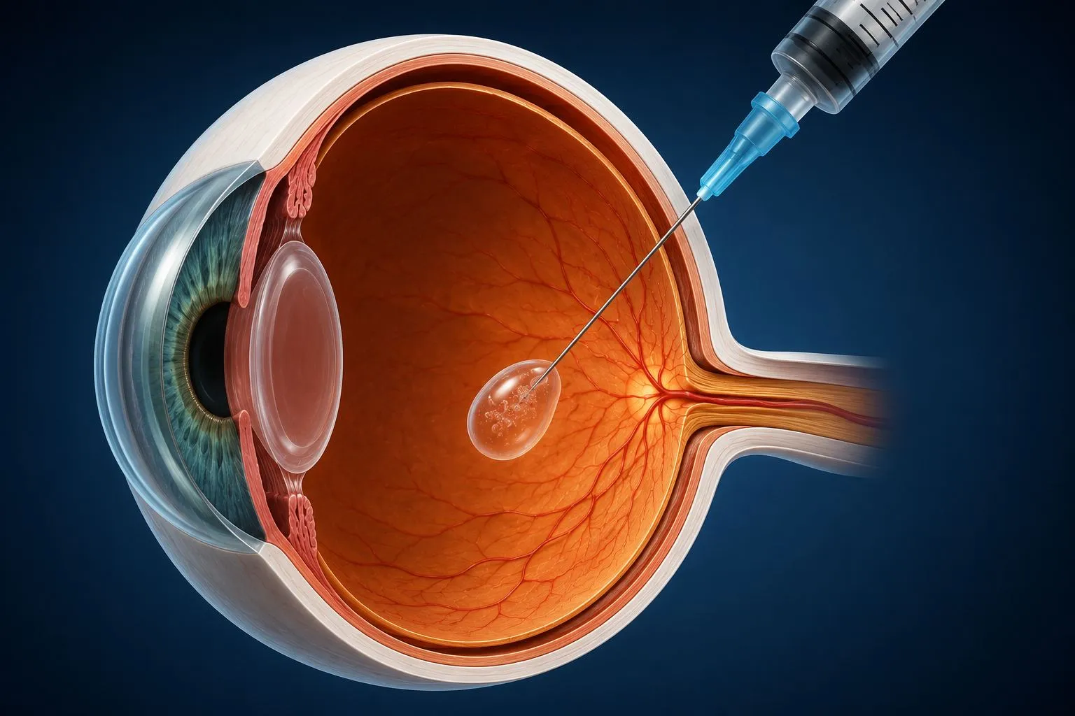 Discover the practice’s dedicated room
Living with diabetes: 4 golden rules for your eyes
Even if your vision seems perfect — the disease is silent.
The targets that protect the retina: HbA1c < 7%, blood pressure < 130/80, LDL cholesterol < 1 g/L — and stopping smoking. Each point of HbA1c gained reduces the risk of complications by a third.
Pregnancy can accelerate retinopathy: close follow-up is essential.
Blurred vision, a spot, distortion: seek care without delay — emergencies are seen on the same day.
Official factsheets from the Société Française d'Ophtalmologie
Written by SFO experts, they describe how each procedure is carried out, and its benefits and risks. Please read them before your consultation.
Eye follow-up for diabetes in Noisy-le-Grand
The Grand Paris Ophtalmologie practice provides screening and treatment of diabetic retinopathy for patients from Noisy-le-Grand and eastern Paris (93, 94, 77): Bry-sur-Marne, Villiers-sur-Marne, Champs-sur-Marne, Neuilly-sur-Marne, Gournay-sur-Marne, Noisiel, Torcy, Chelles, Gagny and Marne-la-Vallée — imaging, laser and injections on site.
Dr Mayer Srour, trained at CHI de Créteil and, and Dr Yaïr Chetrit work in coordination with your diabetologist and your GP (médecin traitant).
Diabetes and vision: your questions
Answers from Dr Mayer Srour and Dr Yaïr Chetrit, ophthalmologists in Noisy-le-Grand.
Why does diabetes damage the eyes?
Chronically excessive blood sugar weakens the small vessels of the retina: they leak, become blocked or grow abnormally. This is diabetic retinopathy — invisible to the patient for a long time.
How often should the eyes be checked?
At least one fundus examination a year, from diagnosis for type 2. More often in cases of known retinopathy, pregnancy or poor blood sugar control.
Are there any symptoms?
Not at first: the disease progresses silently for years. Waiting for vision to drop means treating too late — hence systematic annual screening.
What is diabetic macular oedema?
Fluid that builds up at the centre of the retina because of leaking vessels: it is the leading cause of vision loss in people with diabetes. Detected on OCT, it is treated with injections at the practice.
How is diabetic retinopathy treated?
First, controlling the diabetes (HbA1c, blood pressure, cholesterol). Depending on the stage: retinal laser at the practice and intravitreal injections in cases of macular oedema.
Does retinal laser hurt?
No: it is carried out at the practice, seated, under numbing eye drops with a contact lens. Temporary dazzle is possible after the session.
Which examinations are done at the practice for someone with diabetes?
A complete assessment in one visit: ultra-widefield retinal photography, macular OCT, OCT-angiography without injection, fluorescein angiography if needed — on the technical platform.
Can you go blind because of diabetes?
Retinopathy is the leading cause of blindness in working-age adults — but with regular screening and appropriate treatment, more than 90% of severe vision loss is avoided. Blindness occurs mainly when the disease is discovered too late.
Can retinopathy regress?
In the early stages, excellent blood sugar control can make lesions regress; after laser, abnormal vessels regress in most cases. Advanced lesions, however, are irreversible — hence the importance of early screening.
Is follow-up reimbursed?
Yes: the annual assessment of patients with diabetes is fully covered under the ALD scheme (affection de longue durée, the French long-term illness status that gives full reimbursement by the Assurance Maladie), as are laser and injections.
Related reading
Diabetes? Your annual fundus examination is waiting for you.
A complete assessment in one visit at the Noisy-le-Grand practice.
The Grand Paris Ophtalmologie practice in Noisy-le-Grand provides consultations, screening and surgery for patients from neighbouring towns: