

Ultra-widefield retinal photography
A colour panoramic photograph of the fundus, capturing the retina out to its periphery in a single image. A major asset for screening and monitoring diabetic retinopathy, AMD and peripheral lesions.
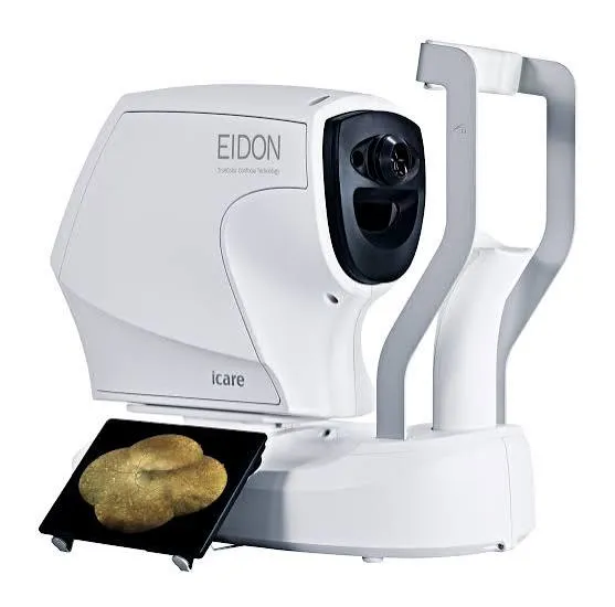
What is ultra-widefield retinal photography?
Retinal photography is a colour photograph of the fundus — that is, of the retina, the membrane lining the back of the eye. The ultra-widefield version captures up to 200°, around 80% of the retinal surface, in a single image.
Where a standard fundus examination shows only the central region, ultra-widefield also documents the periphery of the retina — an area where many lesions hide (tears, diabetes-related lesions), most often without dilating the pupil.
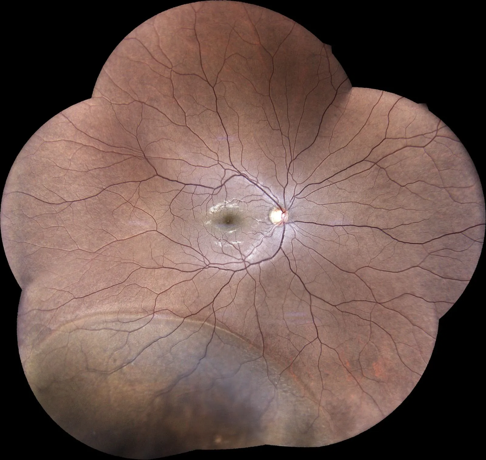
Ultra-widefield or standard retinal photography?
Two ways of photographing the retina — the difference lies in the field covered:
When is it useful?
What happens during the examination?
It could not be simpler: seated, with your chin resting on the device, you look at a target for a few seconds per eye while a brief flash captures the image. No contact, no pain, and most often without dilation — so you leave without being dazzled. The examination takes a few minutes and the images are interpreted immediately by your ophthalmologist, then kept so they can be compared from one visit to the next.
Ultra-widefield retinal photography in Noisy-le-Grand
The Grand Paris Ophtalmologie practice has an ultra-widefield retinal camera, for patients from Noisy-le-Grand and eastern Paris (93, 94, 77): Bry-sur-Marne, Villiers-sur-Marne, Champs-sur-Marne, Chelles, Torcy and Marne-la-Vallée, as well as Le Perreux-sur-Marne, Nogent-sur-Marne, Fontenay-sous-Bois, Vincennes, Saint-Maur-des-Fossés, Créteil, Montreuil and Rosny-sous-Bois. The image is taken and interpreted on site by Dr Yaïr Chetrit or Dr Mayer Srour.
Retinal photography: your questions
Answers from Dr Yaïr Chetrit and Dr Mayer Srour, ophthalmologists in Noisy-le-Grand.
Does the pupil need to be dilated?
Most of the time, no: the ultra-widefield retinal camera obtains a high-quality panoramic image without dilation. You thus avoid several hours of blurred vision and dazzle. Dilation can be added if the periphery needs to be studied closely.
Is it painful?
Not at all: the examination is painless and contactless. You look at a target and a brief flash captures the image — no lasting discomfort.
What is the difference from standard retinal photography?
Ultra-widefield captures up to 200° in a single image, periphery included, whereas a standard fundus photograph shows only the central part.
When is it useful?
Mainly diabetes, AMD, tears and peripheral lesions, high myopia and vascular occlusions — to detect them and follow their progress over time.
Related reading
Have you been prescribed retinal photography?
The examination is carried out at the Noisy-le-Grand practice and interpreted immediately.
The Grand Paris Ophtalmologie practice in Noisy-le-Grand provides consultations, screening and surgery for patients from neighbouring towns: