
AMD — age-related macular degeneration
AMD (age-related macular degeneration) affects central vision after 55. Detected early (OCT, fundus examination), its wet form is treated with intravitreal anti-VEGF injections, which stabilise or improve vision in the majority of cases. At the Noisy-le-Grand practice, for patients from the 93, 94 and 77 areas, Dr Mayer Srour — retina and macula specialist, general secretary of the Fédération France Macula, with more than 15 000 intravitreal injections performed, ranked among the 100 best ophthalmologists in France by Le Point in 2023 — provides screening, treatment and follow-up on site. If you have signs of AMD (wavy lines, a dark spot in the centre, sudden loss of vision), you are seen the same day without an appointment; the OCT and, if an injection is needed, the first injection are carried out the same day, at the practice.
AMD affects central vision after 55. Detected early, it can be monitored and treated. At the Noisy-le-Grand practice, Dr Mayer Srour — macula specialist and General Secretary of the Fédération France Macula — and Dr Yaïr Chetrit provide screening, treatment and follow-up.
AMD: the essentials in 30 seconds
- In an emergency, the same day: signs of AMD = seen without an appointment, OCT and first injection the same day at the Noisy-le-Grand practice (93, 94, 77), by Dr Mayer Srour.
- AMD affects the macula (central vision) after 55; peripheral vision is always preserved.
- Two forms: dry (slow progression) and wet (rapid progression, treatable with anti-VEGF injections).
- Warning signs: straight lines that look wavy, a central dark spot — Amsler grid test on this page.
- The earlier treatment of the wet form starts, the better the vision that is preserved: do not wait to be seen.
What is AMD?
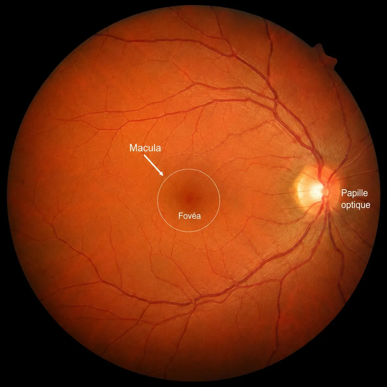
Age-related macular degeneration is a chronic degenerative disease of the retina that begins after 55. It results from overly rapid ageing of the macula — the small central area of the retina (about 2 mm in diameter), right in the optical axis, which transmits 90% of the visual information processed by the brain.
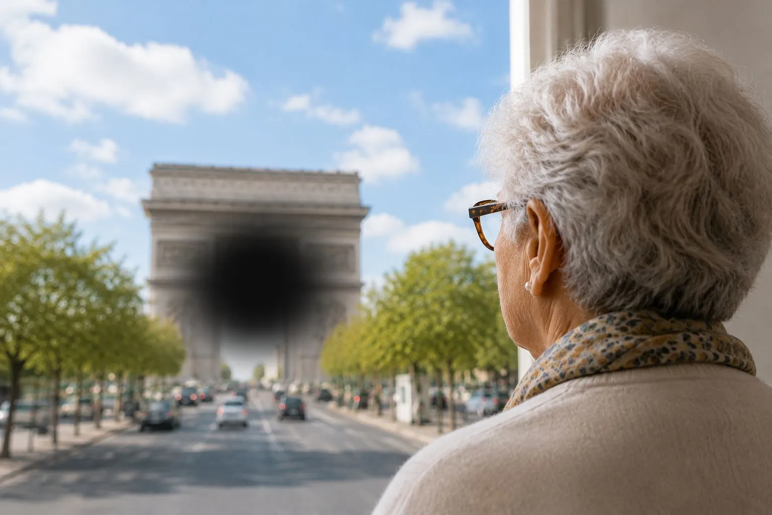
AMD causes a progressive loss of central vision (reading, faces, detail), while usually leaving peripheral vision intact: it never causes total blindness, but it greatly affects daily life if not managed.
A stage precedes it: age-related maculopathy, defined by the presence of drusen on the fundus. Not all drusen carry the same outlook, and the risk of progressing to AMD can be quantified — I have drusen, is it serious?
Symptoms that should alert you
If you notice any of these signs, be seen promptly — the practice sees emergencies the same day. You can also screen yourself with the Amsler grid test below.
The Amsler grid test
A simple test to do at home every week if you are over 55 or have known AMD:
- Wear your usual reading glasses and position yourself 30–40 cm from the screen.
- Cover one eye with your hand, and fix your gaze on the central point with the other.
- While keeping your gaze on the point, observe the rest of the grid.
- Repeat with the other eye.
The two forms of AMD
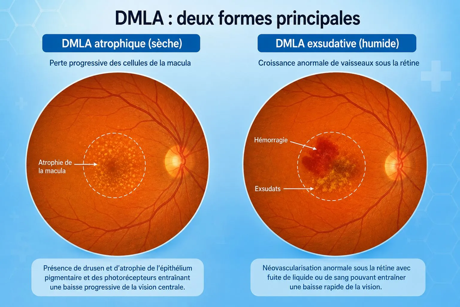
Slow and gradual progression: the cells of the pigment epithelium disappear little by little, and the macula wastes away. Regular follow-up makes it possible to detect in time any change to the wet form.
Abnormal vessels develop at the centre of the macula, which can cause oedema or bleeding and a rapid loss of central vision. It is treated with anti-VEGF injections — hence the importance of a diagnosis without delay.
The AMD assessment with OCT
Macular OCT (optical coherence tomography) is the reference test for AMD: painless, contact-free and without injection, it produces in a few seconds a true microscopic “cross-section” of the retina.
It shows the drusen, the atrophy of the dry form, and above all the signs of the wet form: oedema, cystic spaces, subretinal fluid and neovessels.
Repeated at each check-up, it measures the response to anti-VEGF injections and guides the rhythm of treatment — at the right time, neither too early nor too late.
The practice has a Spectralis OCT (Heidelberg) and an OCT angiography module for a complete assessment in a single visit.
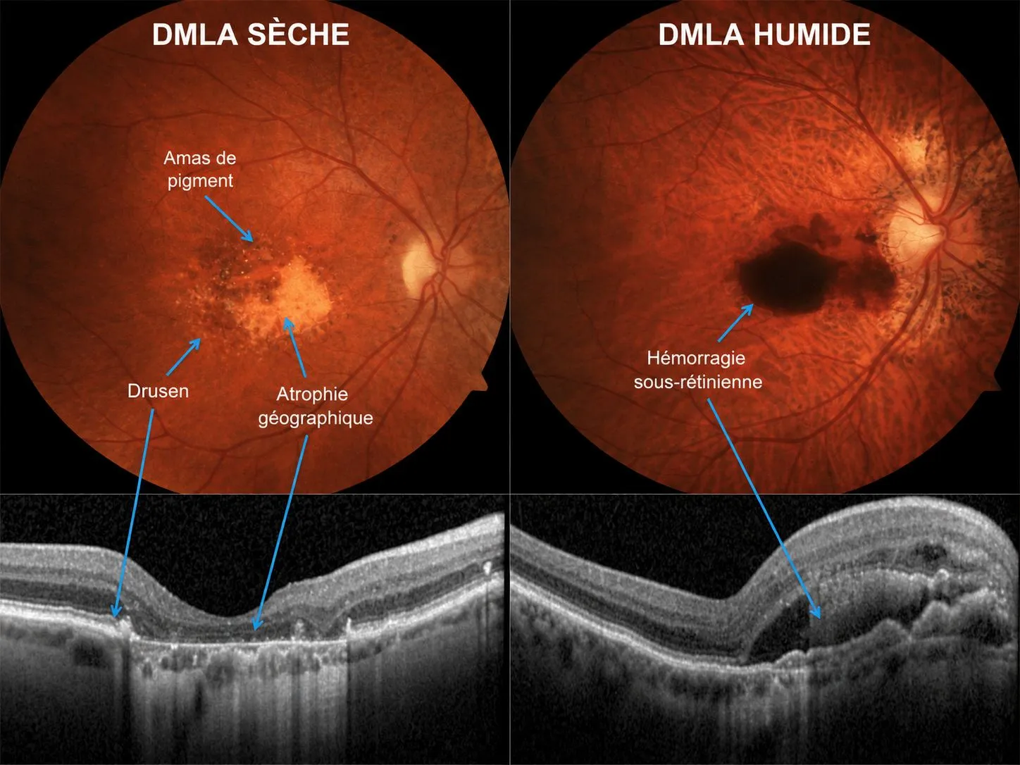
On the left, dry AMD (drusen, geographic atrophy); on the right, the wet form (subretinal haemorrhage) — with the corresponding OCT scans.
Treatments
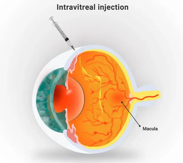
Wet AMD: intravitreal anti-VEGF injections inhibit the growth of abnormal vessels and allow stabilisation, or even regression of the lesions. The injection lasts only a few seconds, under eye-drop anaesthesia, with a very fine needle — performed at the Noisy-le-Grand practice, in a dedicated room with strict aseptic conditions. The rhythm of injections and OCT checks is then personalised.
Dry AMD: no curative treatment yet, but suitable vitamin supplementation may slow progression in some patients, and imaging surveillance (OCT, OCT angiography, ultra-wide-field retinal photography) detects in time any change to the wet form.
AMD explained in video by Dr Srour
Short videos to understand the disease, the injections and good everyday habits.
Find all of Dr Srour’s videos on his YouTube channel: Subscribe to the channel
Polypoidal choroidal vasculopathy: what treatment today?
Dr Mayer Srour answers the question “Polypoidal vasculopathy: which treatment today?” in an interview conducted by Dr Charlotte Rohart, following the scientific day DMLA en pratique (Paris, 18 September 2026).
Biosimilars in AMD: Dr Srour's interview
Dr Mayer Srour answers the question “Is the use of biosimilars inevitable?” at the end of the DMLA en pratique congress (Paris, 12 September 2025).
What a patient with AMD sees
In under 40 seconds, this video shows how AMD changes vision: a dark spot appears at the centre of the image and straight lines look wavy, while vision at the sides is preserved.
The right habit: test each eye regularly with the Amsler grid. If the lines become distorted or an area disappears, be seen promptly — emergencies are seen the same day at the practice.
An intravitreal injection, filmed
Dr Mayer Srour shows in a few seconds how an injection really takes place at the practice — a brief, precise and painless procedure. It should help you arrive more at ease on the day of your injection.
Watch the video on InstagramPolypoidal choroidal vasculopathy (PCV)
Polypoidal choroidal vasculopathy is a particular form of wet AMD, in which the abnormal vessels form small dilatations in a “cluster of polyps” in the choroid, beneath the retina. It can cause pigment epithelial detachments and sometimes substantial haemorrhages. It is more common in people of Asian or African origin, and differs from “classic” wet AMD — hence the importance of recognising it correctly.
OCT and especially OCT angiography now show the polyps and the vascular network with performance close to indocyanine green angiography, in a non-invasive way. Angiography remains the reference in complex cases.
Anti-VEGF injections are the basis of treatment. In PCV, combining them with photodynamic therapy (Visudyne PDT) often gives better regression of the polyps and reduces the number of injections.
Dr Mayer Srour has a particular interest in PCV: he provides diagnosis by multimodal imaging and, when indicated, offers Visudyne PDT — at the practice and in liaison with the reference centres.
AMD: can you have cataract surgery?
Yes. Many patients do not know this: AMD is not a contraindication to cataract surgery. On the contrary, when the drop in vision is also due to cataract, the operation can help you see better. The expected gain depends on the condition of the macula, assessed before the procedure (fundus examination and OCT).
In a patient treated with injections for exudative AMD, the intravitreal injection can, in some cases, be given at the end of the cataract operation: a single visit to the operating theatre and one fewer injection session.
In this video filmed through the operating microscope, you can see the drug spreading in the vitreous, just behind the new implant. Every situation is different: the decision is made case by case with your ophthalmologist.
You are not alone in facing the disease
Low vision rehabilitation & optical aids
When central vision is permanently affected, all is not lost: low vision rehabilitation, carried out with an orthoptist, teaches you to use the remaining peripheral vision (eccentric fixation) to keep reading and stay independent.
Suitable optical aids complete it: illuminated magnifiers, magnifying lenses, video magnifiers, enhanced lighting and optimised contrast. Your ophthalmologist will direct you to the right team (orthoptist, low vision optician).
Association DMLA
Information, a listening ear and mutual support between patients: the association supports people with AMD and their families throughout France.
Low vision: professionals near the practice
Indicative list (directory of ARIBa, the association of low vision professionals, and networks of specialist opticians). Remember to call before travelling.
Specialist opticians: low vision optical equipment (17)
- Optic Maffrand
207 rue Pierre Brossolette, 93160 Noisy-le-Grand · 01 43 03 33 78 - Lissac Bay 2
2-6 Mail Est, 77616 Collégien (Marne-la-Vallée) · 01 64 80 05 72 - Optic 2000 Chelles
2 rue Gustave Nast, 77500 Chelles · 01 60 20 77 96 - Optic 2000 Roissy-en-Brie
1 avenue Ancel de Garlande, 77680 Roissy-en-Brie · 01 60 28 17 53 - Krys Montfermeil
Centre commercial des 7 Îles, 186 avenue Jean Jaurès, 93370 Montfermeil · 01 43 30 56 15 - Krys Rosny-sous-Bois
19/21 rue du Général Gallieni, 93110 Rosny-sous-Bois · 01 45 28 98 49 - Krys Val d'Europe
14 cours du Danube, 77700 Serris · 01 64 63 48 96 - Krys Nogent-sur-Marne
136/138 Grande Rue Charles de Gaulle, 94130 Nogent-sur-Marne · 01 48 73 17 49 - Vision 2000 Nogent
22 boulevard Gallieni, 94130 Nogent-sur-Marne · 01 48 85 15 23 - Vision 2000 Champigny
47 avenue Roger Salengro, 94500 Champigny-sur-Marne · 01 43 83 60 65 - Lissac Vincennes
Rue de Fontenay, 94300 Vincennes · 01 43 98 21 21 - Krys La Varenne
77 avenue du Bac, 94210 La Varenne-Saint-Hilaire · 01 48 83 74 03 - Krys Saint-Maur – Le Lido
95 boulevard de Créteil, 94100 Saint-Maur-des-Fossés · 01 48 83 07 57 - Optique Croix Souris (home visits)
5 rue de la Varenne, 94100 Saint-Maur-des-Fossés · 01 43 97 39 39 - Stef'Optique
36 rue du Moutiers, 94370 Sucy-en-Brie · 01 45 90 12 13 - Lissac Créteil Soleil
Centre commercial Créteil Soleil, 94000 Créteil · 01 49 80 09 79 - Lissac Rivoli
114 rue de Rivoli, 75001 Paris · 01 44 88 44 44
Orthoptists and centres: low vision rehabilitation (7)
- Ms Catherine Koulourath, orthoptist
Low vision assessment and rehabilitation · 5 rue de la République, 93160 Noisy-le-Grand · 01 43 05 55 81 - CHI de Créteil – low vision consultation
Ophthalmology department, 40 avenue de Verdun, 94000 Créteil · 01 45 17 52 30
Fax 01 57 02 29 29 · ophrendezvous@chicreteil.fr (reply within 48 h) - Voir Ensemble – SAMSAH Remora 77
Orthoptist and occupational therapist · 20 rue Pierre-Mendès-France, 77200 Torcy · 01 60 17 09 70 - Centre Simone Delthil – ILVM
Orthoptists and optician · 74 rue Ambroise Croizat, 93200 Saint-Denis · 01 42 35 90 51 - Institut de Réadaptation Visuelle Saint-Louis (IRVSL)
Hôpital national de la vision des Quinze-Vingts · 9 rue Moreau, 75012 Paris - Fondation Rothschild – low vision department
25 rue Manin, 75019 Paris · Book an appointment - Escavie – Cramif
Occupational therapy and home adaptation, visits throughout Île-de-France · 01 40 05 67 51
Official factsheets from the Société Française d'Ophtalmologie
Written by SFO experts, they describe how each treatment is carried out, and its benefits and risks. Please read them before your consultation.
Expertise trained at the world centre for the macula
Dr Mayer Srour trained for 8 years at the Centre Hospitalier Intercommunal de Créteil — a department of worldwide renown for macular disease. He remains attached to it today and continues active research on AMD there, with some thirty publications in international journals. He has performed more than 15 000 intravitreal injections and is among the 100 best ophthalmologists in France in the Le Point 2023 ranking.
General Secretary of the Fédération France Macula, he helps to spread the latest advances among French ophthalmologists. At the practice, you therefore benefit from the most up-to-date protocols — screening, injections and follow-up in Noisy-le-Grand, for all of eastern Paris (93, 94, 77), without going into Paris.
 Interview · RFI radio — Priorité Santé
AMD: your questions
Answers from Dr Mayer Srour, retina and macula specialist, and Dr Yaïr Chetrit, ophthalmologists in Noisy-le-Grand.
Does AMD cause blindness?
No: it affects central vision but usually leaves peripheral vision intact. When managed early, its progression can be slowed, stabilised, or even partially reversed in the wet form.
What are the risk factors?
Age (after 55), a family history of AMD, smoking, and certain genetic and nutritional factors. Regular screening is all the more important if you have several of these factors.
Do the injections hurt? How long do they take?
The injection lasts only a few seconds, under eye-drop anaesthesia, with a very fine needle inserted at a precise and safe point of the eye. It is performed at the practice, in a dedicated room — no hospital stay.
How many injections will I need?
It varies according to the response to treatment: after a loading phase (often 3 injections 4 weeks apart), the rhythm is personalised according to OCT checks, gradually spacing out the injections.
I have signs of AMD: how soon will I be seen?
The same day, without an appointment. Wavy lines, a dark spot in the centre or a sudden drop in central vision are emergencies: come directly to the Noisy-le-Grand practice, Monday to Friday from 8.30 am to 7 pm (emergencies are also seen between 1 pm and 2 pm), or call 01 85 10 57 00. Emergencies are fitted in during the day and are generally seen in under 2 hours.
Dry AMD: is there anything that can be done?
Yes: suitable vitamin supplementation may slow progression in some patients, and imaging follow-up detects in time any change to the wet form, which can be treated. Stopping smoking is strongly recommended.
How often should I do the Amsler test?
Once a week, one eye at a time, if you are over 55 or have known AMD. Any new distortion of the lines or dark spot should prompt you to be seen promptly — the practice sees emergencies the same day.
Which tests are done at the practice for AMD?
Fundus examination, macular OCT (painless, contact-free, 5 minutes), OCT angiography without injection, ultra-wide-field retinal photography and, if needed, angiography with dye — all on the practice's on-site equipment, in a single visit.
Is AMD treatment reimbursed?
Yes: imaging tests and intravitreal injections are procedures covered by the Assurance Maladie (the French national health insurance) under the usual conditions.
How long between diagnosis and the first injection?
No delay if the injection is needed: Dr Mayer Srour performs the OCT, makes the diagnosis and gives the first injection the same day, in the practice's injection room. Subsequent injections are scheduled in weeks (Q4 in the loading phase, then gradual spacing according to the OCT), always on site.
Where can I be seen for AMD near Noisy-le-Grand?
At the Grand Paris Ophtalmologie practice, 67 rue de Malnoue in Noisy-le-Grand (93160). Screening (OCT, fundus examination, OCT angiography), diagnosis and intravitreal injections are all carried out on site, without going through a hospital department. The practice sees patients from Seine-Saint-Denis, Val-de-Marne and Seine-et-Marne, and is five minutes from the A4 motorway and the RER A.
Which ophthalmologist should I see for AMD in eastern Paris?
Dr Mayer Srour is a retina and macula specialist: formerly a hospital practitioner at the retina reference centre of the CHI de Créteil, general secretary of the Fédération France Macula, with more than 15 000 intravitreal injections performed. At the practice, he provides AMD screening, diagnosis, injections and follow-up.
Do I need a referral from another doctor for AMD?
No, you can book an appointment directly. If you have signs suggestive of wet AMD — distortion of straight lines, a central spot, rapid loss of vision — you are seen the same day without an appointment, and the first injection can be given the same day if needed.
Distorted vision? Do not wait.
The earlier wet AMD is treated, the better vision is preserved. The practice sees emergencies the same day.
Related reading
Conditions
All the conditions managed at the practice — click to find out more.
Show all conditions
The Grand Paris Ophtalmologie practice in Noisy-le-Grand provides consultations, screening and surgery for patients from neighbouring towns: