
Polypoidal choroidal vasculopathy
Polypoidal choroidal vasculopathy (PCV) is a disease of the blood vessels of the choroid: an abnormal vascular network ending in “polyp”-like dilations, responsible for detachments and haemorrhages beneath the retina. Close to wet AMD but different from it, it is treated with anti-VEGF injections, alone or combined with photodynamic therapy. Page written by Dr Mayer Srour, first author of the French guidelines.
PCV: the essentials in 30 seconds
- What is it? An abnormal, branching vascularisation of the inner choroid, with aneurysmal dilations (polyps), which can cause a serous or haemorrhagic detachment of the pigment epithelium.
- PCV or AMD? PCV occurs earlier (around age 60), more often in women in Europe, without drusen, on a choroid that is often thick. Freund now classifies it within the pachychoroid spectrum, under the name “type 1 neovascularisation with aneurysms”.
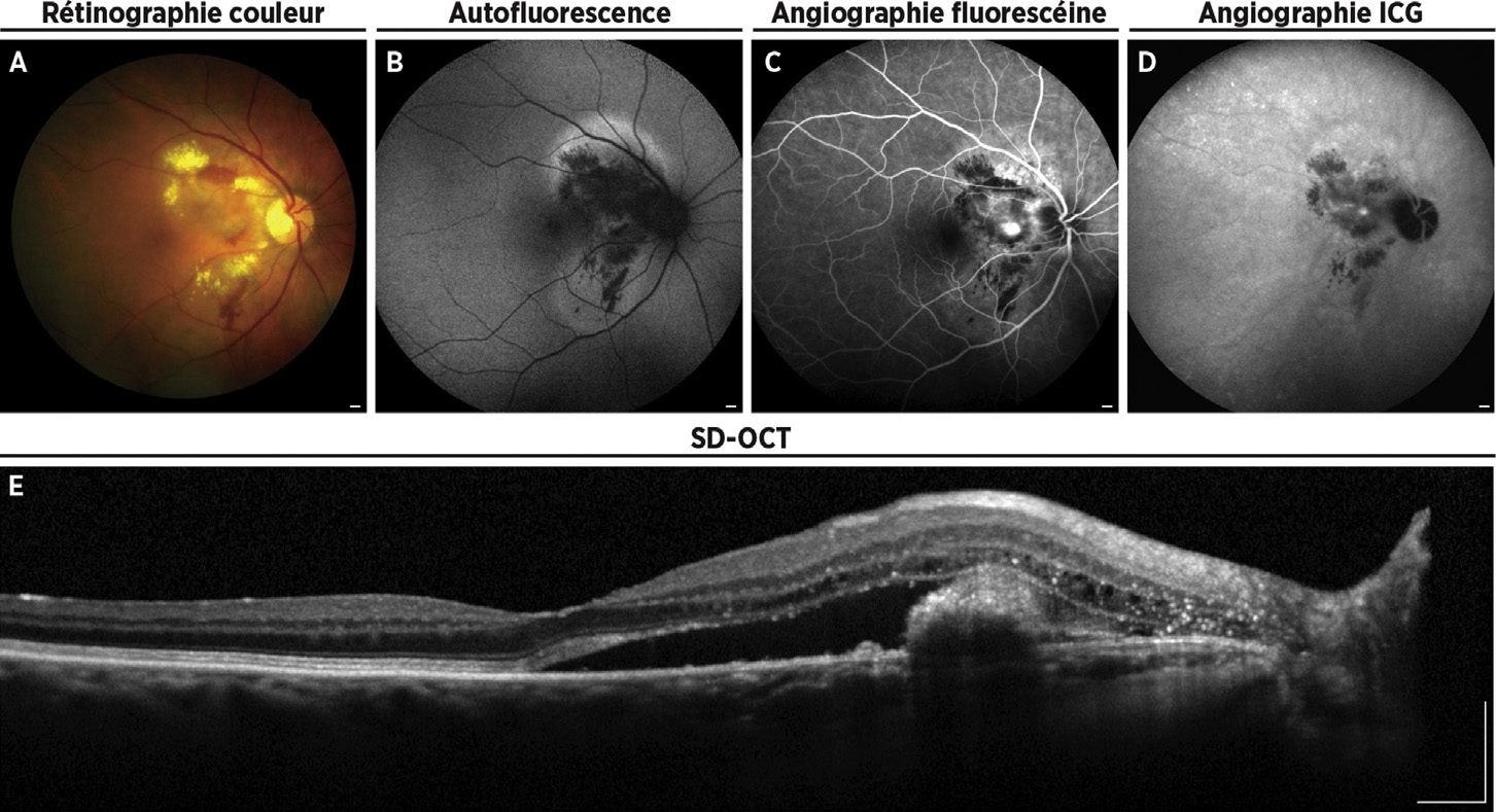
- Diagnosis: multimodal imaging, with indocyanine green angiography (ICG) as the reference; OCT shows the “dome-shaped” or “ogival” detachment of the pigment epithelium typical of the polyp.
- Treatment: first-line, intravitreal anti-VEGF injections as monotherapy, or combined anti-VEGF + verteporfin photodynamic therapy depending on the location of the polyps (guidelines of the Fédération France Macula).
What is polypoidal choroidal vasculopathy?
First described by Yannuzzi in 1982, PCV is an abnormal, branching vascularisation of the inner choroid, associated with aneurysmal vascular dilations — the polyps. These lesions can lead to a serous detachment of the pigment epithelium, a serous retinal detachment and sometimes a true haemorrhagic picture.
Most often idiopathic, it can also be secondary to exudative AMD, to a staphyloma in high myopia, to diffuse retinal epitheliopathy (chronic CSCR) or to a naevus. More recently, Freund has included it in the pachychoroid spectrum and renamed it “type 1 neovascularisation with aneurysms”.
PCV or AMD: what are the differences?
However, the two conditions share risk factors: smoking (risk multiplied by 4 in a Chinese study) and certain genes (CFH, HTRA1, LOC387715), with different frequencies. The answer is therefore not settled: PCV is both close to AMD and a distinct entity in its own right, which justifies a specific therapeutic approach.
How is it diagnosed?
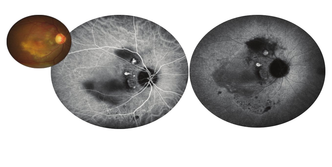
Diagnosis rests on multimodal imaging. The Fédération France Macula recommends indocyanine green angiography (ICG), which remains the gold standard: it shows the abnormal vascular network (branching vascular network) and the polyps as rounded hyperfluorescent dots.
Which treatments?
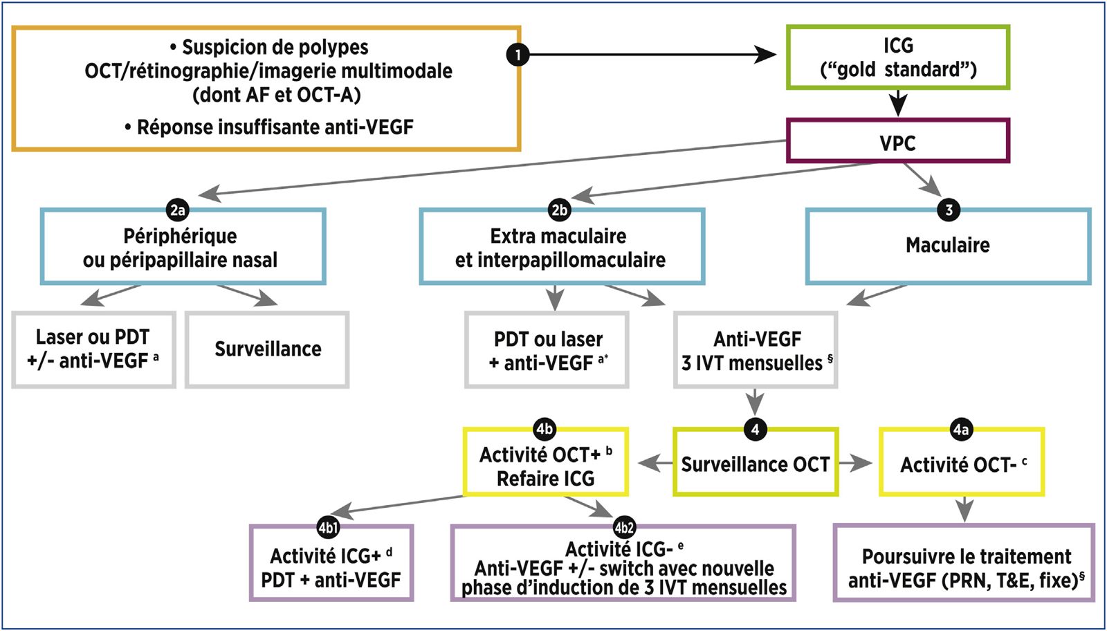
The guidelines of the Fédération France Macula (Srour et al., Journal Français d'Ophtalmologie, 2019), based on analysis of the literature and expert opinion, propose as first-line treatment:
The choice depends on the location of the lesions, visual acuity and response to treatment. A detailed decision algorithm is given in the original article. Follow-up is prolonged, with regular OCT checks: PCV is a chronic, relapsing disease.
Why is it important to identify PCV correctly?
Because PCV responds differently from classic AMD: polyps are sometimes resistant to anti-VEGF alone, and photodynamic therapy, little used in AMD, retains an important place here. A precise diagnosis, with ICG, avoids undertreatment and unnecessary injections. The practice has the full imaging platform required, and Dr Srour coordinated the French guidelines on this condition.
Care in Noisy-le-Grand
Dr Mayer Srour, a retina and macula specialist trained at the Centre Hospitalier Intercommunal de Créteil and author of this publication, sees patients from eastern Paris (93, 94, 77) at the Grand Paris Ophtalmologie practice (67 rue de Malnoue, Noisy-le-Grand, 93). The practice’s equipment allows a complete assessment on site: OCT Spectralis, OCT angiography, angiography, retinal photography, and intravitreal injections are performed at the practice.
Polypoidal choroidal vasculopathy: your questions
Answers from Dr Mayer Srour, ophthalmologist specialising in the retina in Noisy-le-Grand.
Is PCV a form of AMD?
This is debated. It shares risk factors with wet AMD and is treated with anti-VEGF, but it occurs earlier, without drusen, on a thick choroid, and sometimes responds better to combined treatment with photodynamic therapy. Most experts now regard it as an entity within the pachychoroid spectrum, distinct from AMD.
What are the symptoms of PCV?
Loss of vision in one eye, distorted lines, a central spot, sometimes suddenly in the event of a subretinal haemorrhage. Some peripheral PCV remains asymptomatic for a long time.
Is ICG angiography essential?
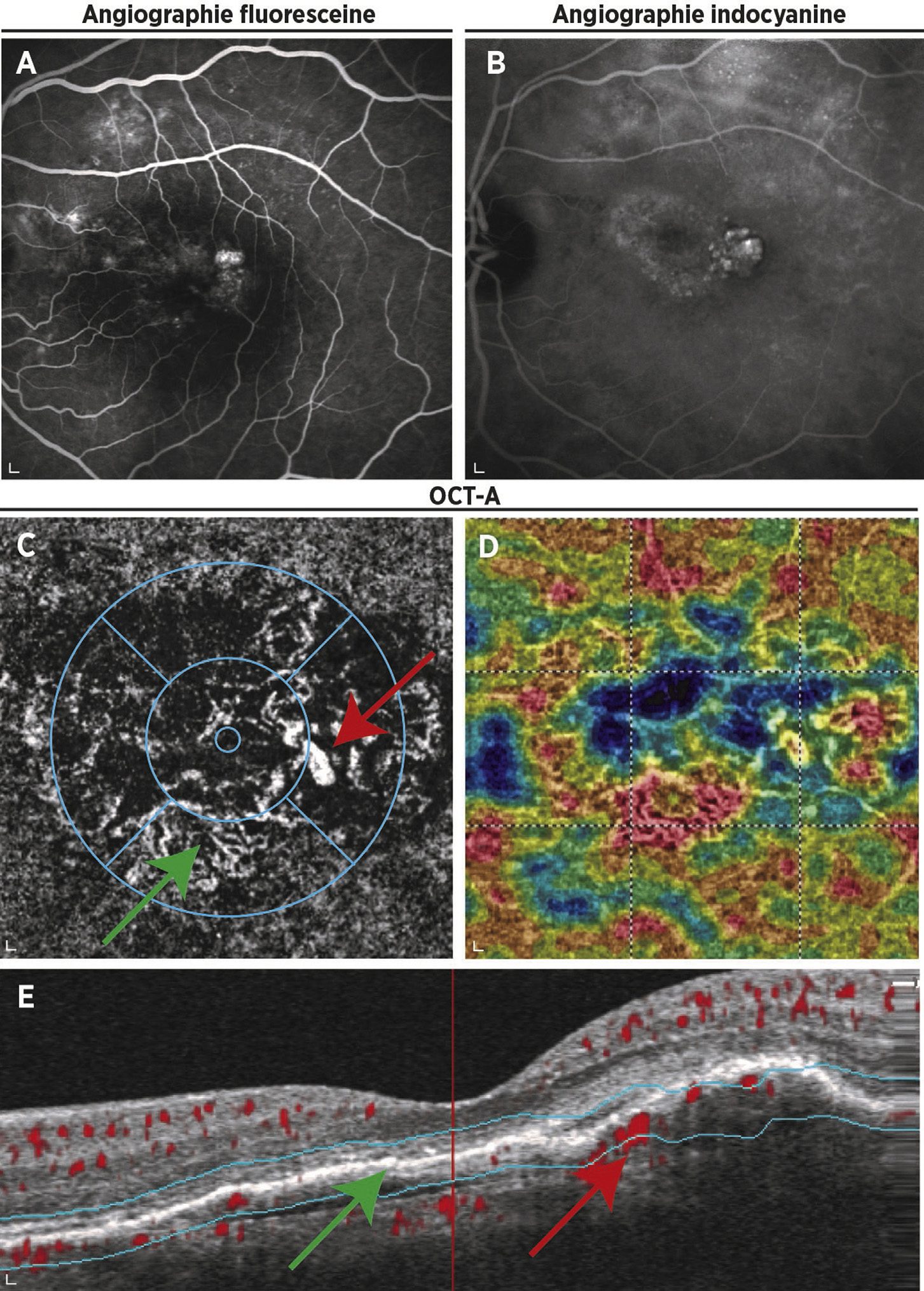
It remains the reference examination for seeing the polyps and the abnormal network. OCT and OCT-angiography often make it possible to strongly suspect the diagnosis; ICG confirms it and guides photodynamic therapy.
What is photodynamic therapy (PDT)?
A two-stage treatment: infusion of a photosensitising drug (verteporfin), then illumination of the lesion with a low-power laser that selectively closes the abnormal vessels. It is done as a day case; sunlight must be avoided for 48 hours.
How many injections will I need?
It depends on each patient. An induction phase of 3 injections at 4-week intervals is usual, then the schedule is adapted to lesion activity on OCT. Combined treatment with PDT may reduce the number of injections needed.
Can PCV be cured?
Polyps can regress completely, particularly after photodynamic therapy, but recurrences are possible. Regular follow-up makes it possible to treat any resumption of activity early and to preserve vision.
Polypoidal choroidal vasculopathy: what treatment today?
Dr Mayer Srour answers the question “Polypoidal vasculopathy: which treatment today?” in an interview conducted by Dr Charlotte Rohart, following the scientific day DMLA en pratique (Paris, 18 September 2026).
How should a polyp be treated? Dr Srour's interview
Dr Mayer Srour answers the question “How should a polyp be treated?” in this interview conducted by Dr Oudy Semoun at the MaculART 2019 congress (Paris).
Related reading
A suspected PCV or one under treatment?
Complete imaging assessment and treatment (anti-VEGF, PDT referral) with Dr Srour, in Noisy-le-Grand.
The Grand Paris Ophtalmologie practice in Noisy-le-Grand provides consultations, screening and surgery for patients from neighbouring towns: