
The pachychoroid
The pachychoroid is an abnormally thick choroid with dilated vessels, visible on OCT. It is not a disease in itself but a background that brings together several macular conditions, from CSCR to neovascular forms. Page written by Dr Mayer Srour based on his publication in Réalités Ophtalmologiques.
Pachychoroid: the essentials in 30 seconds
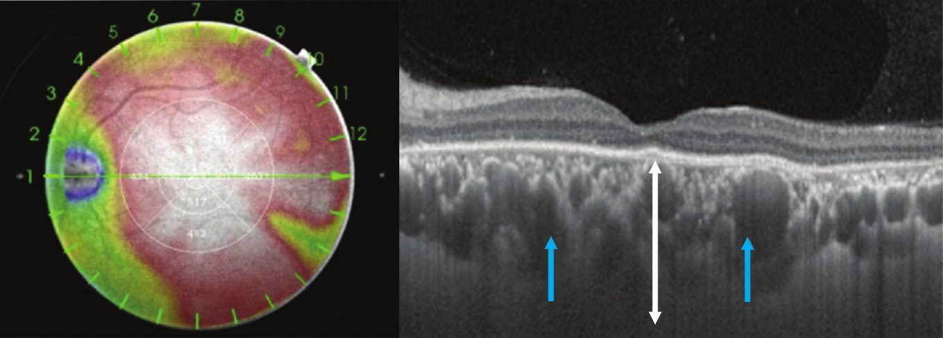
- Definition: diffuse or localised thickening of the choroid (the vascular layer beneath the retina), with large dilated vessels (« pachyvessels ») and hyperpermeability visible on indocyanine green angiography.
- It is not a disease: many people have a thick choroid without any problem. It is considered a disorder when it is accompanied by involvement of the retina or the retinal pigment epithelium.
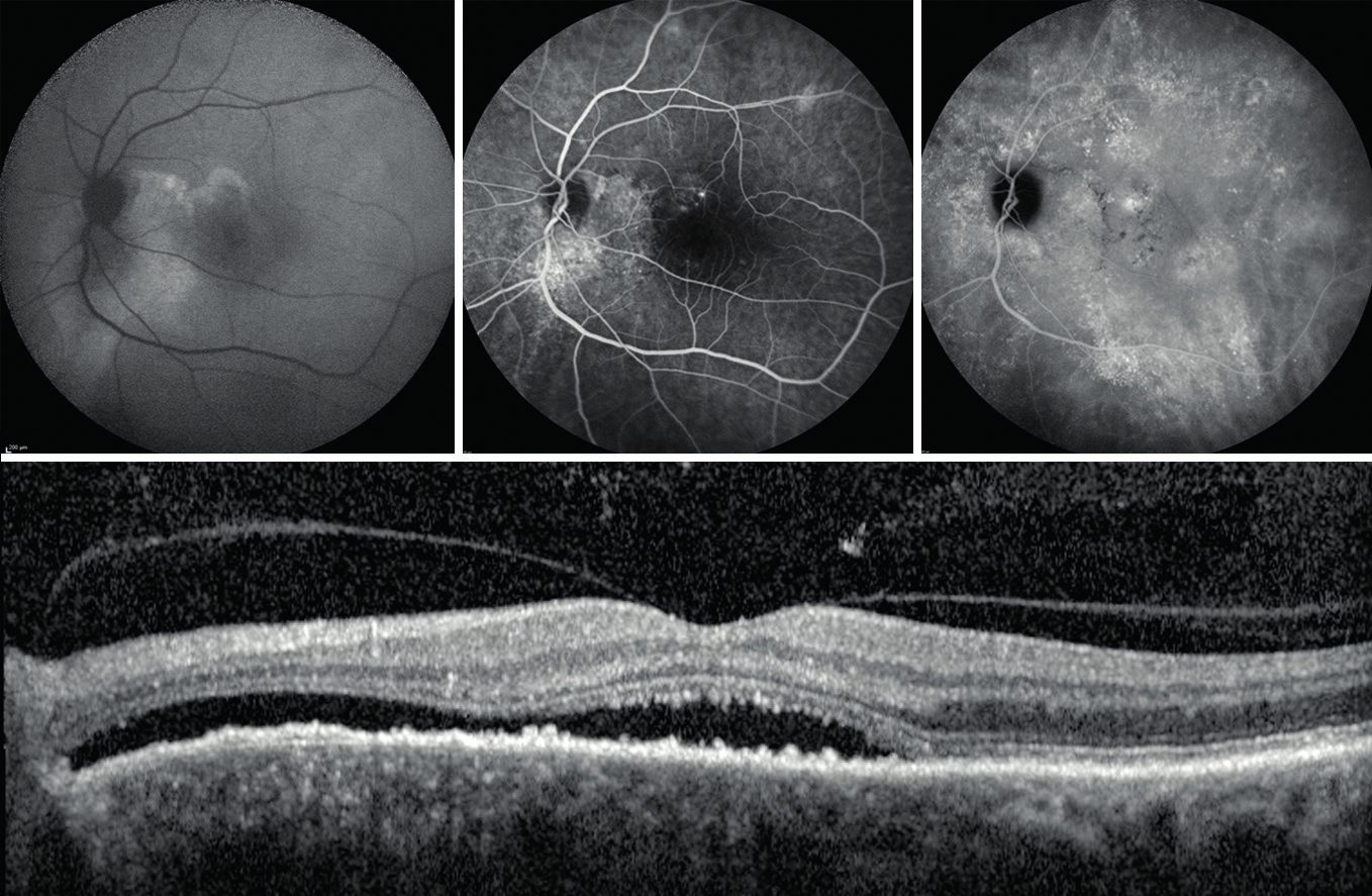
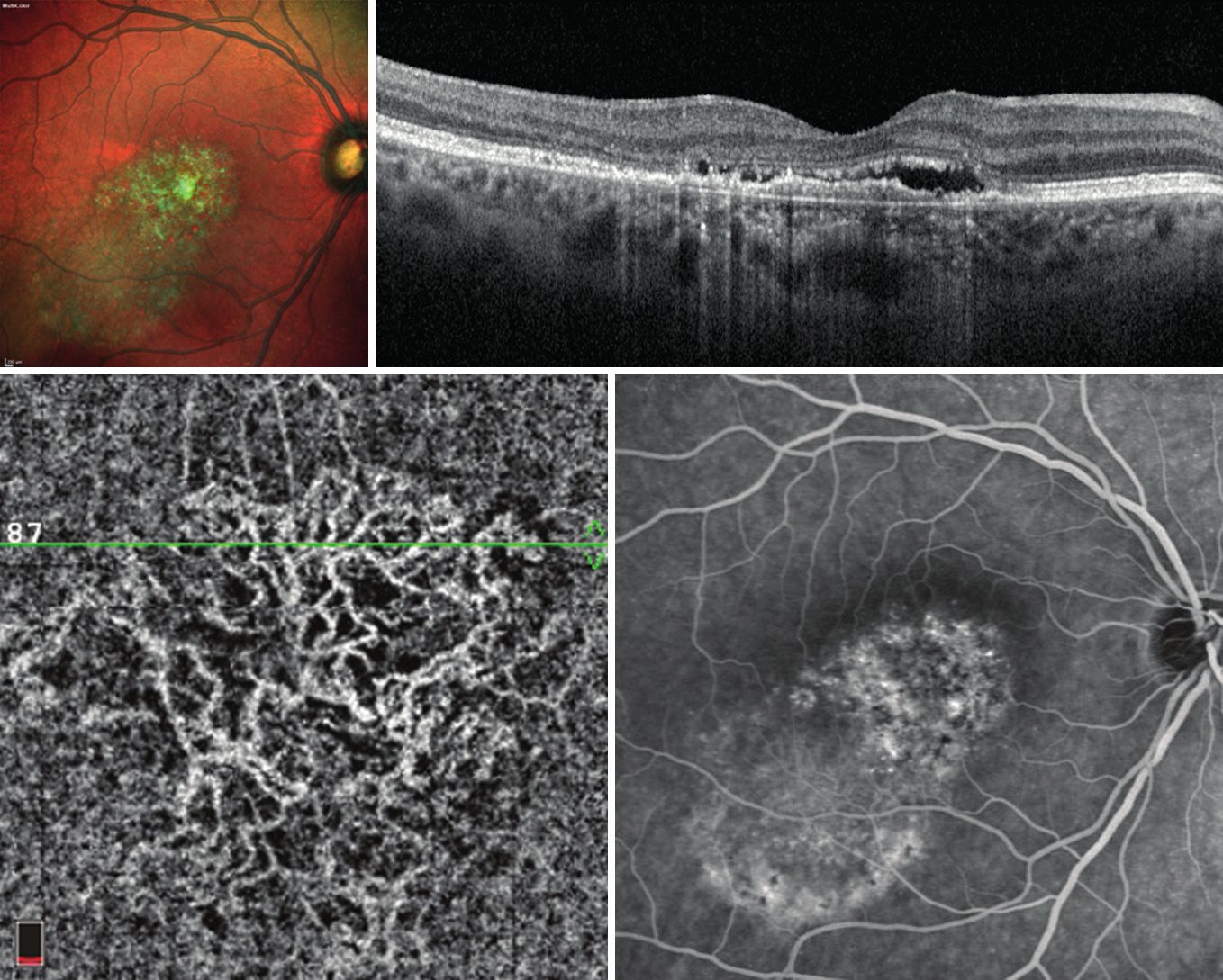
- The forms: non-neovascular — pigment epitheliopathy, peripapillary syndrome, acute or chronic central serous chorioretinopathy (CSCR) — and neovascular — pachychoroid neovasculopathy (PNV) and polypoidal vasculopathy (PCV).
- Treatment: monitoring or laser/photodynamic therapy for non-neovascular forms, anti-VEGF injections ± photodynamic therapy for neovascular forms. OCT angiography is the reference examination for detecting them.
What is the pachychoroid?
The choroid is the vessel-rich layer beneath the retina, which nourishes it. The pachychoroid (from the Greek pachys, thick) was described in 2013 by the team of K. Bailey Freund: it combines a diffuse or focal increase in choroidal thickness, a dilatation of the large vessels of Haller’s layer (pachyvessels) in contact with Bruch’s membrane, and a thinning of the choriocapillaris and of Sattler’s layer.
It is measured with OCT in Enhanced Depth Imaging mode or with Swept-Source, which allows the choroid to be seen in depth. Indocyanine green angiography (ICG) also shows hyperpermeability of the choroidal vessels. Its pathophysiology remains poorly understood: abnormal anastomoses between the draining veins of the choroid (vortex veins), described by Spaide, point towards a disorder of venous congestion.
Non-neovascular forms
All these forms share the same background: a thick choroid with pachyvessels on OCT. CSCR is the most common manifestation.
Neovascular forms
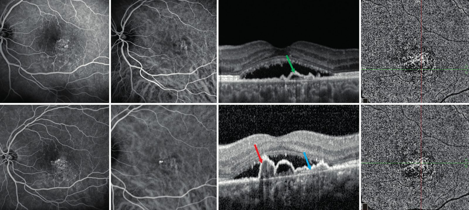
The presence of a pachychoroid is a predictive factor of response to anti-VEGF, and combined anti-VEGF + photodynamic therapy treatment may prove particularly effective in these forms.
How is it diagnosed?
The diagnosis relies on multimodal imaging, fully available at the practice:
Which treatments?
Non-neovascular forms: acute CSCR often resolves spontaneously; stopping corticosteroids and managing stress are essential. If it persists (chronic CSCR), a micropulse laser or half-dose photodynamic therapy (PDT) may be offered to dry up the fluid.
Neovascular forms (PNV, PCV): intravitreal anti-VEGF injections are the first-line treatment, alone or combined with photodynamic therapy depending on the location of the lesions. Regular OCT follow-up makes it possible to adjust the rhythm of the injections.
In all cases, follow-up is long-term: these conditions are relapsing and a non-neovascular form may, over time, be complicated by neovessels.
Pachychoroid and AMD: what are the differences?
Both affect the macula and can lead to neovessels, but AMD occurs on a rather thin choroid, after the age of 60-70, with drusen, whereas conditions in the pachychoroid spectrum often occur earlier, without drusen, on a thick choroid. The distinction matters for treatment: the response to anti-VEGF and the value of photodynamic therapy differ. The two entities probably do, however, share genetic risk factors.
Care in Noisy-le-Grand
Dr Mayer Srour, a retina and macula specialist trained at the Centre Hospitalier Intercommunal de Créteil and author of this publication, sees patients from eastern Paris (93, 94, 77) at the Grand Paris Ophtalmologie practice (67 rue de Malnoue, Noisy-le-Grand, 93). The practice’s equipment allows a complete assessment on site: OCT Spectralis, OCT angiography, angiography, retinal photography, and intravitreal injections are performed at the practice.
The pachychoroid: questions and answers
Answers from Dr Mayer Srour, ophthalmologist specialising in the retina in Noisy-le-Grand.
Is the pachychoroid serious?
Not in itself: an isolated thick choroid is simply an anatomical variant. It is its complications — fluid beneath the retina (CSCR), neovessels, polyps — that can reduce vision. Hence the value of OCT follow-up when a pachychoroid background is identified.
What are the symptoms?
Often none. When fluid accumulates beneath the macula: blurred or darkened vision in one eye, distorted straight lines (metamorphopsia), objects seen smaller (micropsia), a central grey spot. A sudden drop in vision should prompt a quick consultation.
Who is affected?
CSCR typically affects men aged 30 to 50, of an anxious temperament or under stress, working night shifts, or taking corticosteroids (tablets, sprays, creams, infiltrations). Neovascular forms (PNV, PCV) occur rather after 50-60.
Can relapses be prevented?
Triggering factors can be limited: avoid corticosteroids in all forms when possible, regulate sleep, manage stress. An OCT check at the slightest symptom allows early treatment.
What is the role of OCT angiography?
It is the examination that has changed management: it detects, without injection, Type 1 neovessels hidden beneath a flat detachment of the pigment epithelium, often invisible on conventional angiography. It is performed at the practice, during the consultation.
Is the treatment permanent?
No: conditions in the pachychoroid spectrum progress in flare-ups. Treatment dries the retina and protects vision, but regular follow-up remains necessary to detect a relapse or the appearance of neovessels.
Related reading
Distorted vision or a central spot?
Complete macular assessment (OCT, OCT angiography, angiography) at the practice in Noisy-le-Grand, with Dr Srour.
The Grand Paris Ophtalmologie practice in Noisy-le-Grand provides consultations, screening and surgery for patients from neighbouring towns: