

Corneal transplant
When the cornea is too distorted or clouded to be corrected any other way, transplant (keratoplasty) replaces the damaged part with a clear graft. Modern techniques often replace only a single layer of the cornea, for safer recovery.
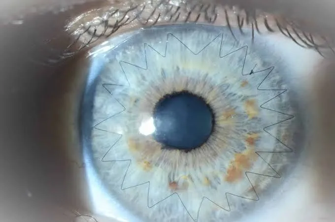
Corneal transplant: the essentials in 30 seconds
- Indicated when the cornea has permanently lost its transparency or shape (advanced keratoconus, dystrophies, scars).
- Modern techniques often replace only the diseased layer (lamellar grafts): faster recovery.
- As the cornea has no blood supply, rejection is less common than with other transplants — but follow-up remains essential.
- Complete assessment at the practice and referral to the appropriate procedure.
What is a corneal transplant?
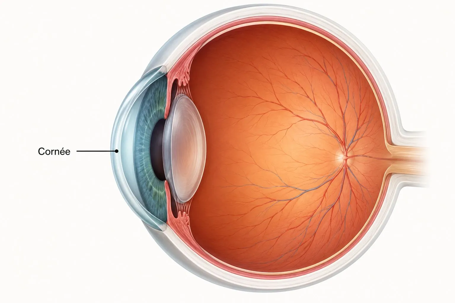
The cornea is the clear lens at the front of the eye. To see well, it must remain clear and regular. When a disease or injury makes it too distorted or opaque, vision falls and cannot be restored with glasses or contact lenses.
A corneal transplant, or keratoplasty, then consists of replacing the damaged part with a clear graft taken from a donor. The cornea is made of several layers: depending on which one is affected, the surgeon replaces either its full thickness or only the diseased layer — a modern, safer approach.
When is a transplant offered?
A transplant is considered when vision can no longer be improved by glasses or contact lenses, or when the cornea is under threat. The main indications:
The different techniques
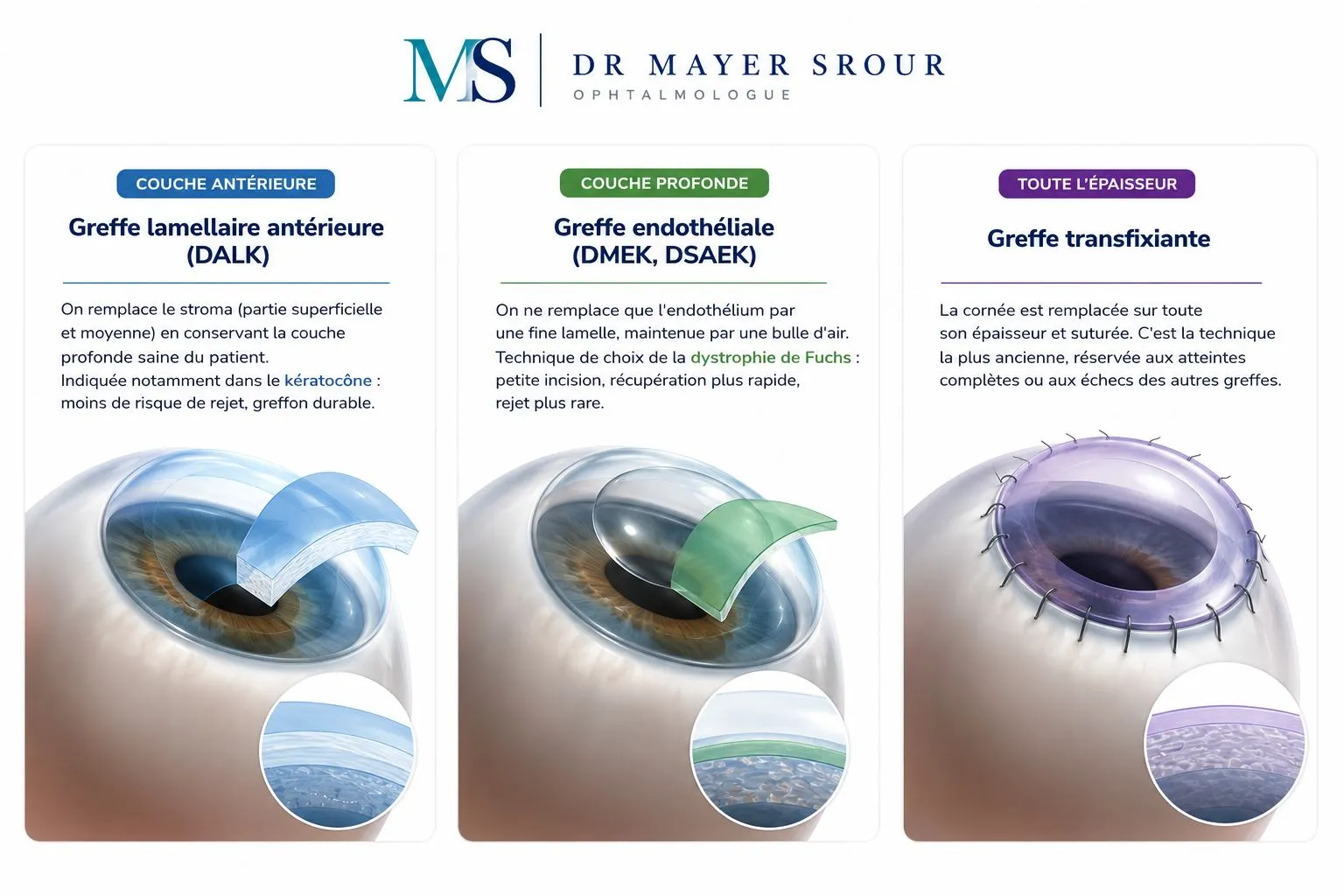
Surgery has evolved greatly: rather than systematically replacing the whole cornea, the aim today is to change only the diseased layer and preserve the healthy layers. The surgeon chooses the technique best suited to each case.
How is the transplant carried out?
A transplant requires some organisation, because it depends on the availability of a graft. The main stages:
Follow-up & the risk of rejection
Corneal transplantation is a well-established procedure with generally good results. Vision returns gradually: the cornea takes several weeks to become clear again, and final adjustment (suture removal, correction) may take several months. Regular follow-up, especially in the first year, is essential.
A red, painful, watering eye, reduced vision or sensitivity to light should prompt urgent consultation. Caught in time, early rejection is most often treated by stepping up corticosteroid eye drops. This is why it is so important never to space out check-ups or stop treatment without medical advice.
Corneal transplant in Noisy-le-Grand
At the Grand Paris Ophtalmologie practice, Drs Chetrit and Srour determine the indication for a corneal transplant, carry out the pre-operative assessment and provide post-operative follow-up on site, in liaison with a surgical centre, for patients from Noisy-le-Grand and east Paris (93, 94, 77): Bry-sur-Marne, Villiers-sur-Marne, Champs-sur-Marne, Chelles, Torcy and Marne-la-Vallée, as well as Le Perreux-sur-Marne, Nogent-sur-Marne, Fontenay-sous-Bois, Vincennes, Saint-Maur-des-Fossés, Créteil, Montreuil and Rosny-sous-Bois. Follow-up is provided by Dr Yaïr Chetrit or Dr Mayer Srour.
Corneal transplant: your questions
Answers from Dr Yaïr Chetrit and Dr Mayer Srour, ophthalmologists in Noisy-le-Grand.
Is a living donor needed?
No. The graft comes from a deceased donor, via a cornea bank. Retrieval, storage and allocation are strictly regulated to ensure the safety and quality of the graft.
How long does it take to recover vision?
Recovery is gradual. The cornea takes several weeks to become clear again, and final adjustment (suture removal, correction) may take several months, especially after a penetrating graft.
Is a corneal transplant painful?
The operation is done under anaesthetic (local or general), without pain. In the following days, mild discomfort or a gritty sensation is possible, relieved by eye drops.
What are the signs of rejection?
A red, painful, watering eye, reduced vision or sensitivity to light. They require urgent consultation: caught in time, rejection is most often treated by stepping up eye drops.
Can both eyes be grafted at the same time?
No: when both eyes need a graft, the second is operated on some time after the first (generally after about a year), once the first graft has stabilised.
Does a graft last a lifetime?
Often for many years. Modern lamellar techniques extend the life of the graft; in case of wear or rejection, a new transplant remains possible.
Related reading
Has a corneal transplant been mentioned to you?
Indication, pre-operative assessment and follow-up at the Noisy-le-Grand practice, with Drs Chetrit and Srour.
The Grand Paris Ophtalmologie practice in Noisy-le-Grand provides consultations, screening and surgery for patients from neighbouring towns: