

Retinal vein occlusion
A vein in the retina becomes blocked: blood backs up and vision drops suddenly in one eye. This is an emergency that needs prompt care. At the Noisy-le-Grand practice, Dr Mayer Srour — a specialist in vascular diseases of the retina — and Dr Yaïr Chetrit provide diagnosis, injections and laser on site.
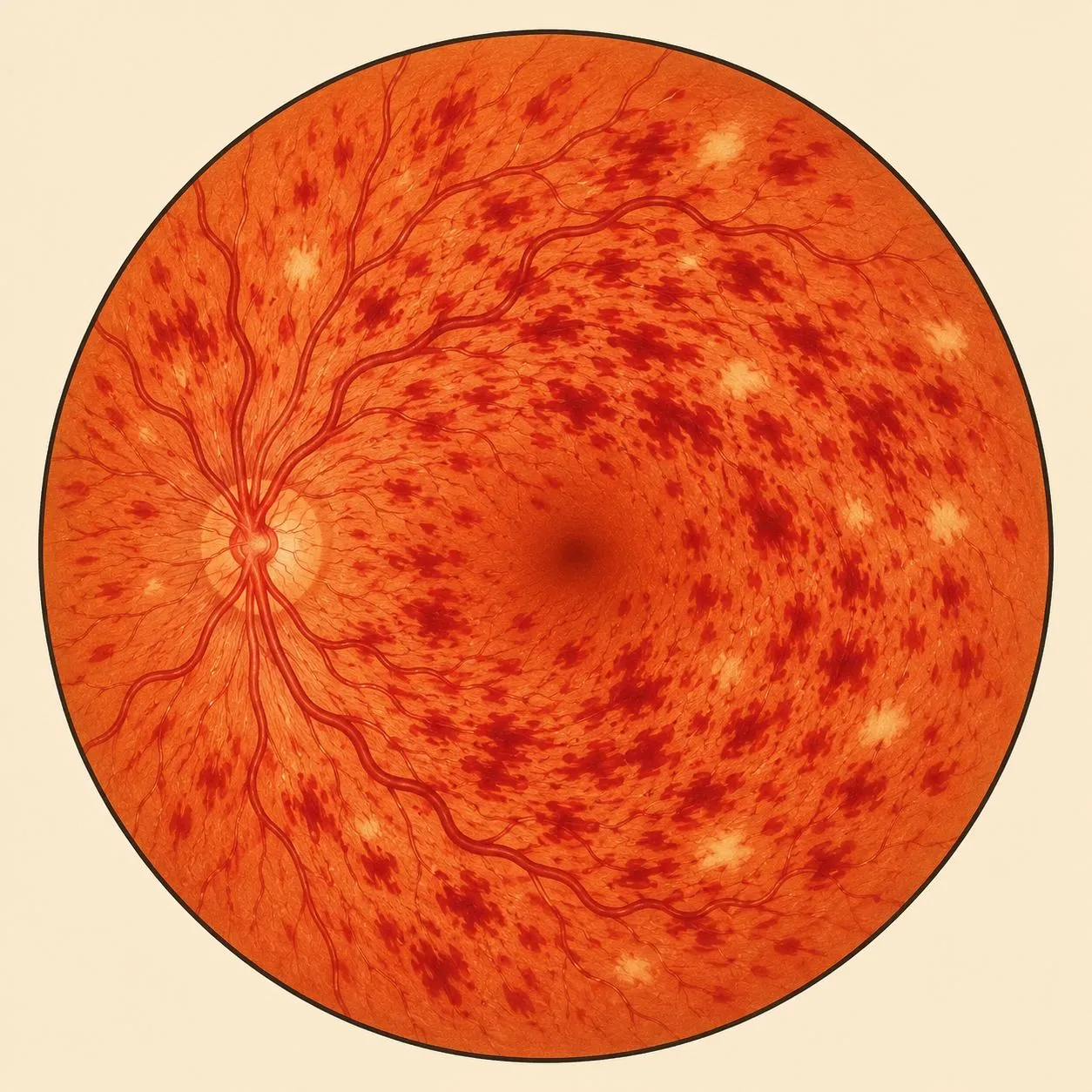
Vein occlusion: the essentials in 30 seconds
- Sudden, painless loss of vision in one eye only — often linked to high blood pressure after the age of 60.
- The resulting macular oedema can be treated effectively with intravitreal injections.
- A cardiovascular check-up with your GP (médecin traitant) is part of the care.
- Regular OCT follow-up at the practice to adjust the interval between injections (in weeks).
What is a vein occlusion?
The retina is nourished by arteries and drained by veins. When a vein becomes blocked (most often by a clot), blood can no longer drain away: it backs up and stagnates in the retina, causing haemorrhages and a build-up of fluid at the centre of vision — macular oedema, the main cause of the loss of vision.
Unlike diabetic retinopathy, vein occlusion occurs suddenly and generally affects one eye only. It is the second most common cause of retinal vascular disease after diabetes, favoured by high blood pressure and age.
The two types of occlusion
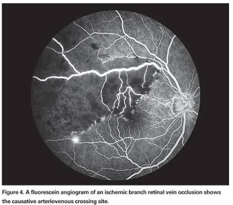
Depending on the vein involved, the occlusion affects the whole retina or only one sector — and depending on the remaining circulation, it is oedematous or ischaemic. These distinctions guide the prognosis and treatment:
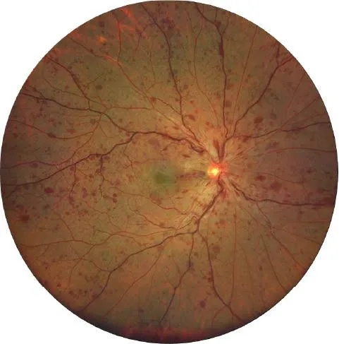
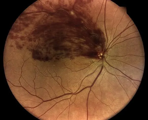
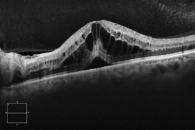
Symptoms & risk factors
The typical symptom is a sudden, painless drop in visual acuity in one eye (sudden loss of vision), or blurred and distorted vision that develops over a few hours to a few days. There is neither pain nor a red eye. Any sudden loss of vision in one eye calls for prompt consultation.
Vein occlusion is favoured by cardiovascular risk factors. Identifying and treating them helps protect the other eye:
Diagnosis, in a single visit
The fundus examination shows characteristic dilated veins and haemorrhages. The assessment is completed in a single visit on the practice’s technical platform:
A general check-up (blood pressure, blood sugar, lipid profile), in liaison with your GP (médecin traitant), systematically completes the care.
Treatments
The foundation: controlling risk factors. Blood pressure, blood sugar and cholesterol are monitored with your GP (médecin traitant) — this is what protects the retina and the other eye in the long term.
For the eye itself, two key treatments are carried out directly at the practice, adapted to the type of occlusion:
Argon laser photocoagulation: in a session at the practice, seated at the slit lamp, under anaesthetic eye drops with a contact lens. Reserved for ischaemic forms — it treats the non-perfused areas of the retina and prevents the development of abnormal vessels and neovascular glaucoma.
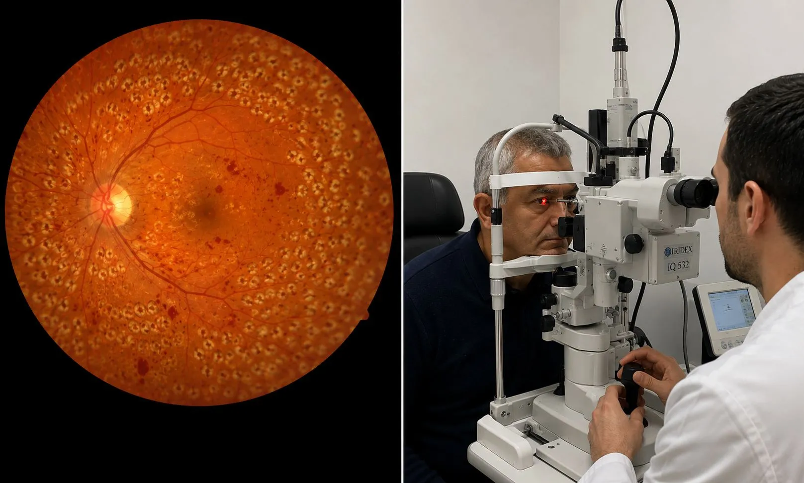 Discover the practice’s laser equipment
Anti-VEGF or corticosteroids against macular oedema: this is the first-line treatment. It takes a few seconds, under anaesthetic eye drops, in the practice’s dedicated room — no hospital stay. A loading phase is followed by spaced-out intervals according to OCT checks.
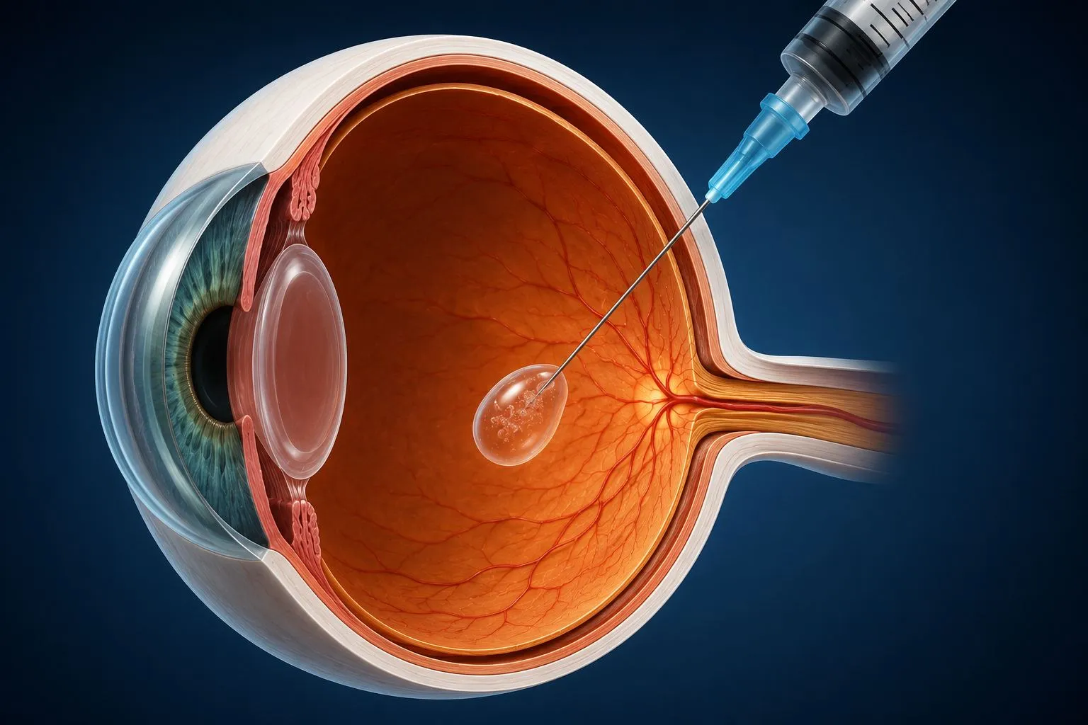 Discover the practice’s dedicated room
When treated early, retinal vein occlusion often recovers well. Everything depends on the speed of treatment and control of blood pressure: this is what protects your vision and your other eye in the long term.
Neovascular glaucoma
In ischaemic forms, the poorly supplied retina can produce abnormal vessels (new vessels) that block the drainage of fluid from the eye. The pressure then rises suddenly: this is neovascular glaucoma, an emergency that threatens vision. Regular screening can prevent it.
Vein occlusion: good habits
Any sudden loss of vision in one eye calls for a prompt examination: the earlier the treatment, the better the recovery.
High blood pressure is the leading risk factor. Treating it protects the other eye and prevents recurrence.
Macular oedema is treated over the long term: sticking to the schedule of injections and OCT checks determines the visual outcome.
Blurred vision, a spot, distortion: seek care without delay — emergencies are seen on the same day.
Official factsheets from the Société Française d'Ophtalmologie
Written by SFO experts, they describe how each treatment is carried out, and its benefits and risks. Please read them before your consultation.
Retinal vein occlusion in Noisy-le-Grand
The Grand Paris Ophtalmologie practice provides diagnosis and treatment of vein occlusion for patients from Noisy-le-Grand and eastern Paris (93, 94, 77): Bry-sur-Marne, Villiers-sur-Marne, Champs-sur-Marne, Neuilly-sur-Marne, Gournay-sur-Marne, Noisiel, Torcy, Chelles, Gagny and Marne-la-Vallée — imaging, injections and laser on site.
Dr Mayer Srour, trained at the CHI de Créteil and a specialist in vascular diseases of the retina, and Dr Yaïr Chetrit work in coordination with your GP (médecin traitant) and your cardiologist.
Good to know: the Association DMLA (a French AMD patient association) and other patient associations for retinal diseases provide information and support to people with retinal vascular conditions.
Vein occlusion: your questions
Answers from Dr Mayer Srour and Dr Yaïr Chetrit, ophthalmologists in Noisy-le-Grand.
What is a retinal vein occlusion?
The blockage of a vein in the retina: blood can no longer drain away, it backs up and causes haemorrhages and macular oedema. A distinction is made between central retinal vein occlusion (CRVO) and branch retinal vein occlusion (BRVO).
What are the symptoms?
A sudden, painless loss of vision in one eye, or blurred vision that develops over a few hours to a few days. No pain and no red eye — but you must be seen quickly.
What are the causes?
Mostly after the age of 50: high blood pressure, cholesterol, diabetes, smoking and glaucoma. In young people, a clotting disorder is looked for.
How is it diagnosed?
Fundus examination, macular OCT, OCT angiography and angiography — to measure the oedema and distinguish the oedematous from the ischaemic form. Everything is done at the practice.
How is vein occlusion treated?
The reference treatment for macular oedema is intravitreal injection of anti-VEGF (or corticosteroids). Laser is reserved for ischaemic forms. Blood pressure control is essential.
What is the difference between the oedematous and ischaemic forms?
The oedematous form (the most common) responds well to injections, with a good prognosis. The ischaemic form carries a risk of abnormal vessels and neovascular glaucoma: close monitoring and laser.
Can vision be recovered?
Yes in many cases, especially if treatment is started early: injections reduce the oedema and vision often improves. Recovery depends on the type of occlusion and how early it is treated.
Is the other eye at risk?
The risk of the other eye being affected is increased. Controlling your blood pressure and your cardiovascular risk factors is the best prevention, together with your GP (médecin traitant).
Is treatment reimbursed?
Yes: consultation, imaging (OCT, angiography), injections and laser are covered by the Assurance Maladie (the French national health insurance).
Related reading
Sudden loss of vision? Be seen quickly.
Diagnosis and treatment of vein occlusion at the Noisy-le-Grand practice — emergencies seen on the same day.
The Grand Paris Ophtalmologie practice in Noisy-le-Grand provides consultations, screening and surgery for patients from neighbouring towns: