

The Spectralis OCT: the retina to the micron
The Spectralis is the tomograph that performs our OCT scans of the macula and optic nerve: cross-sections of the retina accurate to a few microns, essential for monitoring AMD, glaucoma and diabetic retinopathy.
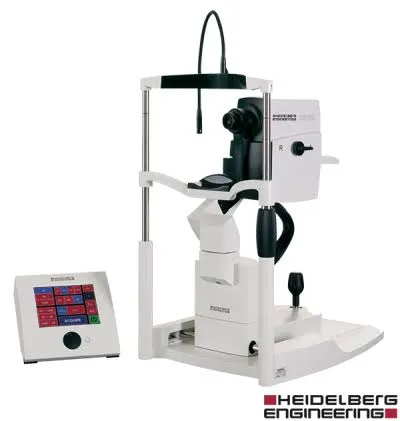
What is the Spectralis, and what is it used for?
The Spectralis is an optical coherence tomograph (OCT): it uses light to produce virtual cross-sections of the retina, layer by layer, like a scanner — but with no X-rays and no contact. The resolution reaches a few microns: each of the layers of the retina, the macula, the optic disc and the optic nerve fibres can be distinguished.
It is the central examination of the modern retina: it detects oedema, fluid under the retina, a membrane, a macular hole or thinning of the optic nerve fibres well before vision is affected.
Why it is a benchmark in its category
The Spectralis’s signature feature is its TruTrack eye-tracking: a second camera constantly follows the micro-movements of your eye and locks the OCT scan onto the retina. The result: sharp images even if the eye moves, and above all a follow-up mode that repositions each scan at exactly the same location as at the previous visit.
For a chronic disease, this is decisive: from one check to the next, the machine compares the very same micron of retina — the slightest recurrence of fluid in AMD treated with injections, or the finest loss of fibres in glaucoma, is detected reliably and not attributable to a difference in positioning.
When the practice uses it
What it means for you
Spectralis OCT: your questions
Answers from Dr Yaïr Chetrit and Dr Mayer Srour, ophthalmologists in Noisy-le-Grand.
What is the difference between OCT and a fundus examination?
The fundus examination looks at the surface of the retina; OCT cuts through it in depth, layer by layer. The two are complementary: one sees the extent, the other the structure.
Does the examination require dilation?
Most often no: the Spectralis images the retina through a normal pupil. Dilation is sometimes useful for certain analyses — the doctor will tell you in that case.
How often does an OCT need to be repeated?
It depends on the disease: at every check for AMD treated with injections, once or twice a year for stable glaucoma. The frequency is set by your ophthalmologist.
Related reading
Have you been prescribed an OCT?
The Spectralis is installed at the Noisy-le-Grand practice: the examination and its interpretation take place on site, on the same day.
The Grand Paris Ophtalmologie practice in Noisy-le-Grand provides consultations, screening and surgery for patients from neighbouring towns: