01 85 10 57 00 67 rue de Malnoue, 93160 Noisy-le-Grand
4,9 960 reviews Open Mon–Fri 8:30–13:00 · 14:00–19:00 LinkedIn Instagram
Grand Paris Ophtalmologie Book an appointment
! Eye emergency Same-day care, no appointment needed — Mon–Fri 8:30–19:00 (including between 13:00 and 14:00) Call 01 85 10 57 00 ! Eye emergency Same-day care, no appointment needed — Mon–Fri 8:30–19:00 (including between 13:00 and 14:00) Call 01 85 10 57 00 ! Eye emergency Same-day care, no appointment needed — Mon–Fri 8:30–19:00 (including between 13:00 and 14:00) Call 01 85 10 57 00 ! Eye emergency Same-day care, no appointment needed — Mon–Fri 8:30–19:00 (including between 13:00 and 14:00) Call 01 85 10 57 00 ! Eye emergency Same-day care, no appointment needed — Mon–Fri 8:30–19:00 (including between 13:00 and 14:00) Call 01 85 10 57 00 ! Eye emergency Same-day care, no appointment needed — Mon–Fri 8:30–19:00 (including between 13:00 and 14:00) Call 01 85 10 57 00
The practice

State-of-the-art technical facilities

Imaging · Examinations · Lasers · Injections

Screening, diagnosis, treatment and follow-up of all eye conditions, in one place — without multiple journeys or delays.

10 examinations
of imaging and testing + 3 lasers on site
Everything on site
examinations, lasers and injections at the practice
The same day
examinations carried out at the consultation if needed
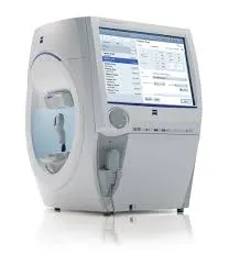
Visual field test at the practice Slit-lamp examination Ophthalmic diagnostic equipment
1 · The 10 examinations 2 · Therapeutic lasers 3 · Injection room (IVT)
1

Examinations carried out at the practice

Retinal angiography
Photographs of the back of the eye after injection of a dye (fluorescein or indocyanine), to visualise the state of the retinal vessels. Useful in retinal vein occlusions, diabetic retinopathy, AMD, retinal inflammation and certain tumours — to make the diagnosis, assess severity and guide treatment.
Learn more about retinal angiography →
Angle OCT (Anterion Analyser)
A latest-generation Swept Source OCT that explores the anterior segment in high definition: corneal topography, biometry of the lens and anterior chamber, iridocorneal angles. Indicated before cataract surgery, when there is a risk of angle-closure glaucoma, or to screen for corneal disease.
Quick and painless · contact lenses removed during the examination · no visual discomfort afterwards
Biometry
High-precision optical measurements of the eye, taken in a few seconds while you look at a light — they make it possible to calculate the made-to-measure implant before cataract surgery.
Painless, without dilation · in the pre-operative assessment, the associated dilated fundus examination means you should not drive afterwards
Visual field test
An assessment of everything you perceive while looking at a fixed point: it shows how the optic nerve is working, from the retina to the visual pathways of the brain. A key examination in the follow-up of glaucoma.
Seated in front of a dome, in the dark · a buzzer in hand to signal each light stimulus you see · requires good concentration
OCT (Macular / Optic nerve)
Optical Coherence Tomography studies the retina, optic nerve and cornea in very precise cross-sections, using a light wave. Measurements are compared with a normative database and with your previous examinations — essential for the diagnosis and follow-up of AMD, diabetes and glaucoma.
Seated, chin on a chin rest · painless, no contact · most often without dilation (OCT Spectralis)
OCT Angiography
Non-invasive analysis, without injection of dye, of the retinal vascular network, in 2 or 3 dimensions: looking for abnormalities of retinal or choroidal perfusion, in particular in AMD, and monitoring of injection treatments (anti-VEGF). Often supplemented, if needed, by angiography with dye.
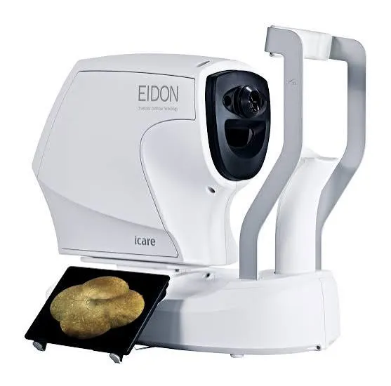
Ultra-Widefield Retinal Photography
A colour photograph of the retina (Eidon device), centred on the macula, the optic nerve or any peripheral lesion. It provides a reliable comparison from one visit to the next: follow-up of patients with diabetes, screening for early signs of AMD, monitoring of a naevus.
Quick and painless · short flash · dilation sometimes necessary (avoid driving afterwards)
Corneal topography
A camera rotating through 360° generates maps of the cornea’s curvature, thickness and relief. It is the reference examination for screening and follow-up of keratoconus, and is essential for the pre-operative assessment of refractive surgery and for contact lens fitting.
No contact with the eye · no discomfort after the examination
UBM Ultrasound
Ultrasound Bio Microscopy: a very high-frequency, non-invasive imaging technique of the anterior segment — cornea, iris, lens and ciliary processes. It visualises the iridocorneal angle to assess glaucoma, screen for a tumour, investigate an anterior chamber abnormality or plan surgery.
B-Mode Ultrasound
Ultrasound of the posterior segment: cross-sections of the whole length of the eye and measurements of the globe, to explore the back of the eye when the media are cloudy — intravitreal haemorrhage, dense cataract, corneal opacity — follow diseases and prepare for surgery.
2

Therapeutic lasers

Three lasers make it possible to treat directly at the practice, without hospitalisation, under eye-drop anaesthesia:

YAG laser
Secondary cataract (capsulotomy) and prevention of acute glaucoma (iridotomy).
SLT laser
Lowers eye pressure in glaucoma — as first-line treatment or in addition to eye drops.
Argon laser
Sealing of retinal tears and treatment of diabetic retinopathy.
Everything about medical lasers
3

The room dedicated to intravitreal injections

The practice has a specially equipped room for intravitreal injections (IVT), carried out under strict aseptic conditions: treatment of wet AMD, diabetic macular oedema and retinal vein occlusions.

Your injections are given on site, without hospitalisation, by the doctor who follows you — with the control OCT in the same place, at every stage of treatment.

Why it matters

Everything in one place, from screening to treatment

An abnormal examination? Imaging is done straight away. A laser to schedule? It is carried out on site. Injections? The dedicated room is next to the consulting room. Fewer delays, fewer journeys — faster and safer care.

Book an appointment

A complete technical facility in Noisy-le-Grand

Few community practices have this kind of equipment: patients from Noisy-le-Grand, Seine-Saint-Denis (93), Val-de-Marne (94) and Seine-et-Marne (77) — Bry-sur-Marne, Villiers-sur-Marne, Champs-sur-Marne, Chelles, Torcy and all of Marne-la-Vallée — have access to imaging and treatments usually reserved for hospital centres, without going to Paris. All in the hands of Dr Yaïr Chetrit and Dr Mayer Srour.

Related reading

Speciality
Medical laser
YAG, SLT and Argon: treatments at the practice.
Speciality
Retinal diseases
AMD, diabetes, glaucoma: follow-up by imaging.
Speciality
Cataract surgery
Biometry for a made-to-measure implant.

Have you been prescribed an examination?

OCT, visual field, topography… everything is done at the practice in Noisy-le-Grand.

Book an appointment on Doctolib 01 85 10 57 00
Our equipment in detail

The practice’s devices, one by one

Each device has its own page: what it does, why it is a reference, and what it concretely changes for you.
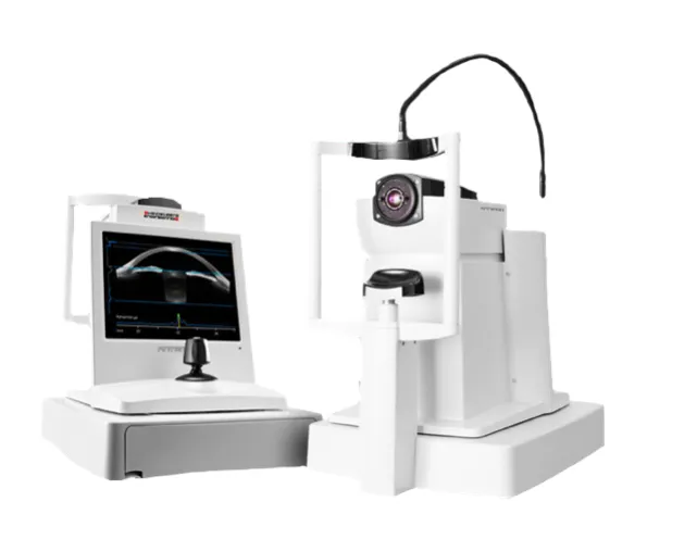
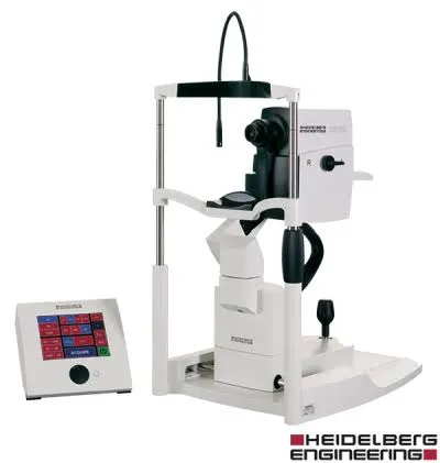
Anterion — Heidelberg Engineering
Biometry, topography and anterior segment imaging: six examinations in a single, non-contact device.
Learn more
OCT Spectralis — Heidelberg
The retina to the micron, with eye-tracking: the reference tomograph for AMD, glaucoma and diabetes.
Learn more
HFA3 — Zeiss
HFA3 — Zeiss
The Humphrey visual field, the world standard for glaucoma: shorter with SITA Faster, GPA progression follow-up.
Learn more
Eidon — iCare
Eidon — iCare
The retina photographed in true colours out to the periphery, most often without dilation.
Learn more
OCT Angiography module
The retinal vessels mapped without dye injection, repeatable at every visit.
Learn more
Page written and reviewed by Dr Mayer Srour, ophthalmic surgeon in Noisy-le-Grand (Grand Paris Ophtalmologie). Last updated: September 2026.