Floaters: those threads drifting in front of your eyes, should you be worried?
What are they? Should you see a doctor? Are there treatments? The carousel published on Instagram by Dr Mayer Srour, to browse here image by image, with the text of each page.


Swipe, or use the arrows. The text of each image is reproduced below.
The carousel, image by image
The text of each carousel page, to read at your leisure or to find a specific piece of information.
Those threads drifting in front of your eyes: should you be worried?
Floaters: what the science says. What are they? Should you see a doctor? Are there treatments?
The shadow of an ageing gel
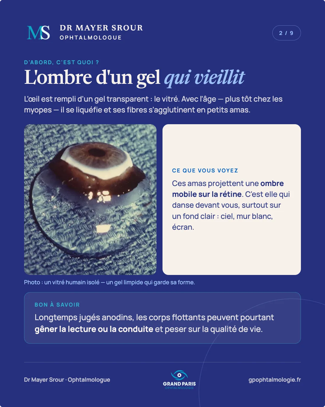
The eye is filled with a clear gel: the vitreous. With age — earlier in people with myopia — it becomes liquid and its fibres clump together into small clusters.
What you see: these clusters cast a moving shadow on the retina. This is what dances in front of you, especially against a light background: sky, white wall, screen.
Good to know: long considered harmless, floaters can nevertheless interfere with reading or driving and affect quality of life.
Threads, dots, cobwebs…
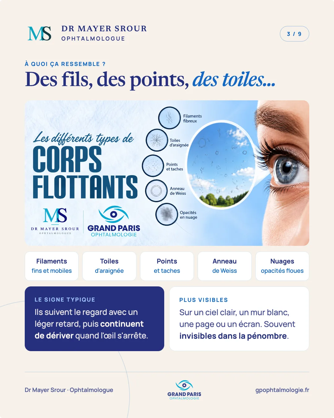
The different types of floaters: fine, mobile filaments, cobwebs, dots and spots, Weiss ring, clouds (blurry opacities).
The typical sign: they follow your gaze with a slight delay, then keep drifting after the eye stops.
More visible against a bright sky, a white wall, a page or a screen. Often invisible in dim light.
The vitreous detaches
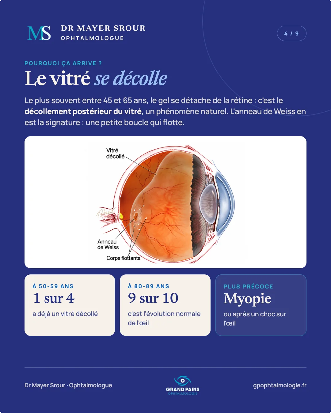
Most often between the ages of 45 and 65, the gel detaches from the retina: this is posterior vitreous detachment, a natural phenomenon. The Weiss ring is its signature: a small floating loop.
At 50-59, 1 person in 4 already has a detached vitreous; at 80-89, 9 in 10: this is the normal evolution of the eye. It occurs earlier in people with myopia, or after a blow to the eye.
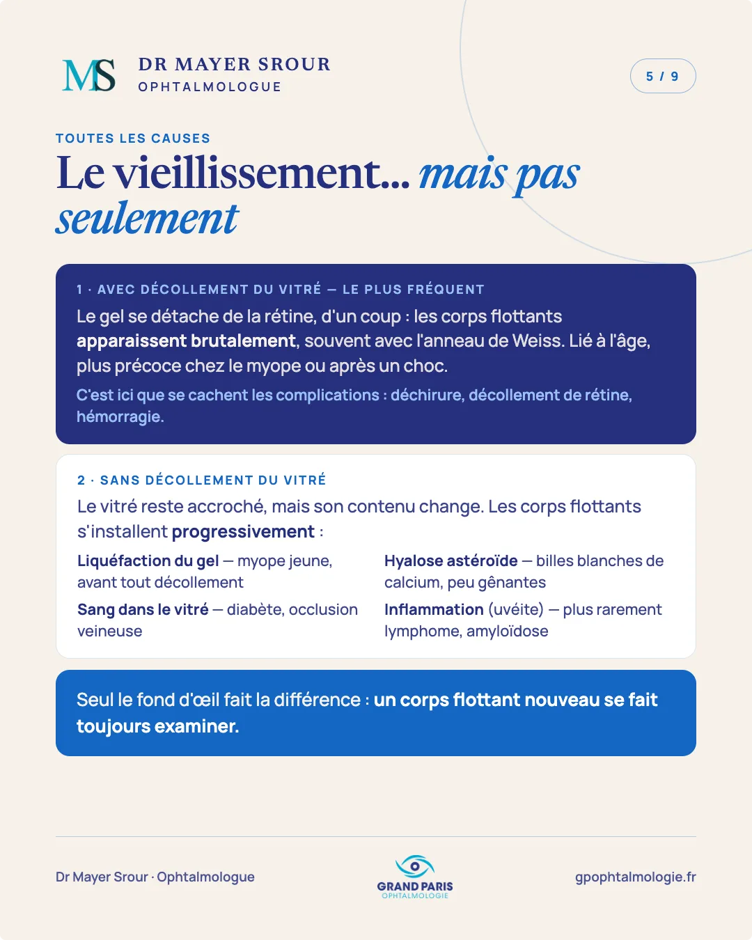
Ageing… but not only
1 · With vitreous detachment — the most common: the gel detaches from the retina all at once; floaters appear suddenly, often with a Weiss ring. Age-related, earlier in people with myopia or after a blow. This is where complications can hide: tear, retinal detachment, haemorrhage.
2 · Without vitreous detachment: the vitreous stays attached, but its contents change; floaters develop gradually. Liquefaction of the gel (young person with myopia, before any detachment), asteroid hyalosis (white calcium beads, rarely troublesome), blood in the vitreous (diabetes, vein occlusion), inflammation (uveitis) — more rarely lymphoma, amyloidosis.
Only a fundus examination can tell the difference: a new floater should always be examined.
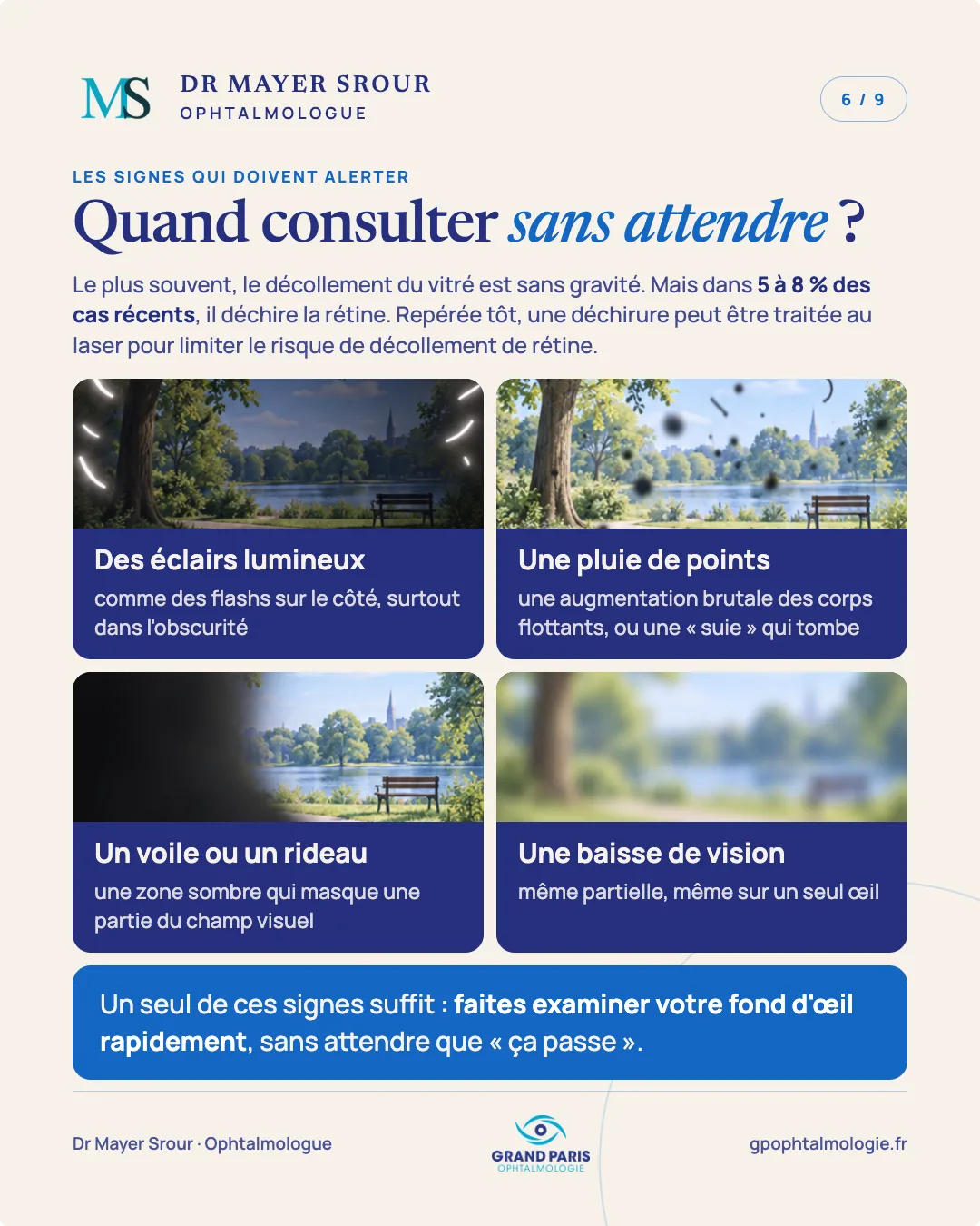
When to see a doctor without delay?
Most often, vitreous detachment is harmless. But in 5 to 8% of recent cases, it tears the retina. If found early, a tear can be treated with laser to limit the risk of retinal detachment.
The signs that should alert you: flashes of light (like flashbulbs at the side, especially in the dark); a shower of dots (a sudden increase in floaters, or a falling “soot”); a veil or a curtain (a dark area hiding part of the visual field); a loss of vision, even partial, even in one eye only.
A single one of these signs is enough: have your fundus examined quickly, without waiting for it to “go away”.
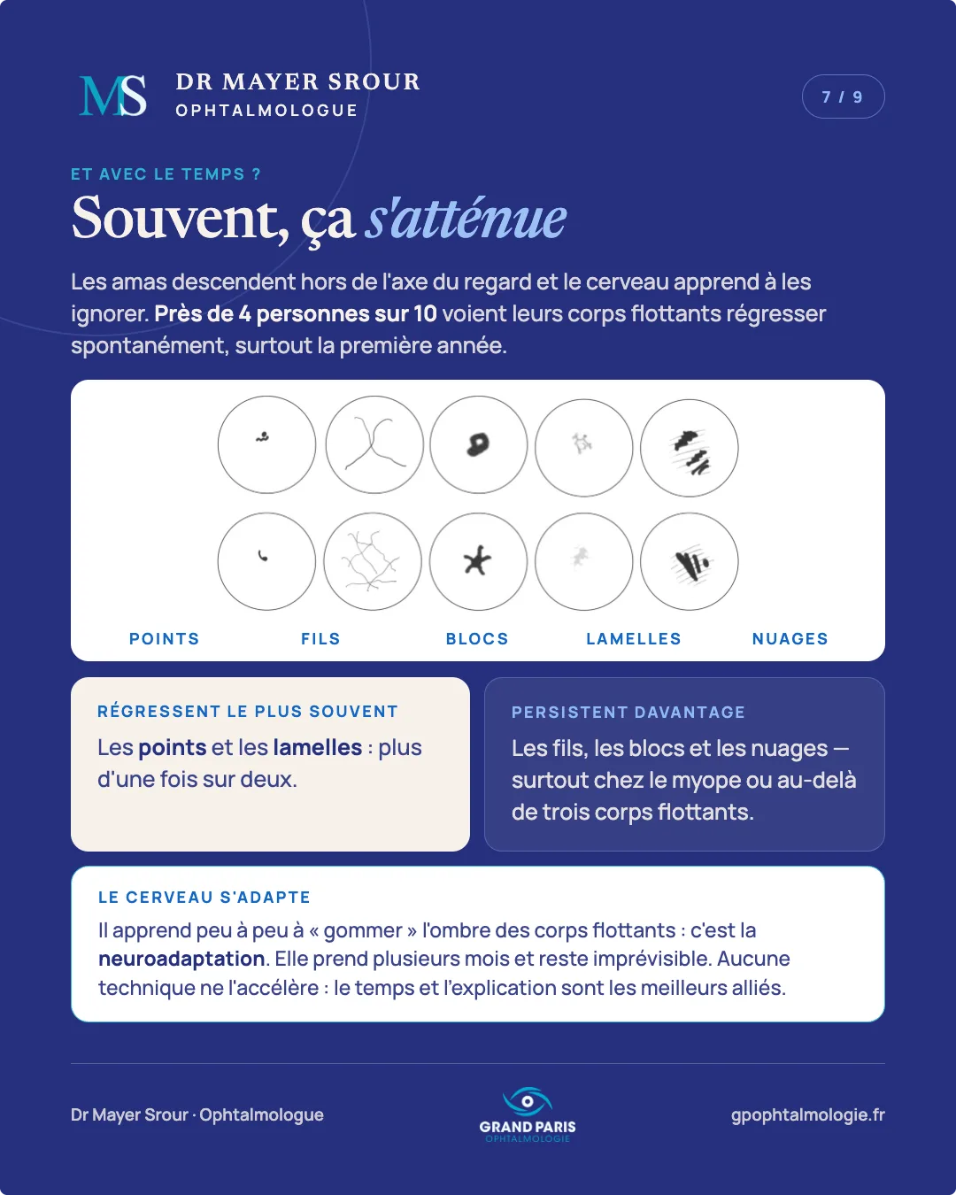
Often, it fades
The clusters sink out of the line of sight and the brain learns to ignore them. Nearly 4 in 10 people see their floaters regress spontaneously, especially in the first year.
Most often regress: dots and flakes — more than one time in two. More likely to persist: threads, blocks and clouds, especially in people with myopia or with more than three floaters.
The brain adapts: it gradually learns to “erase” the shadow of floaters, which is called neuroadaptation. It takes several months and remains unpredictable. No technique speeds it up: time and explanation are the best allies.
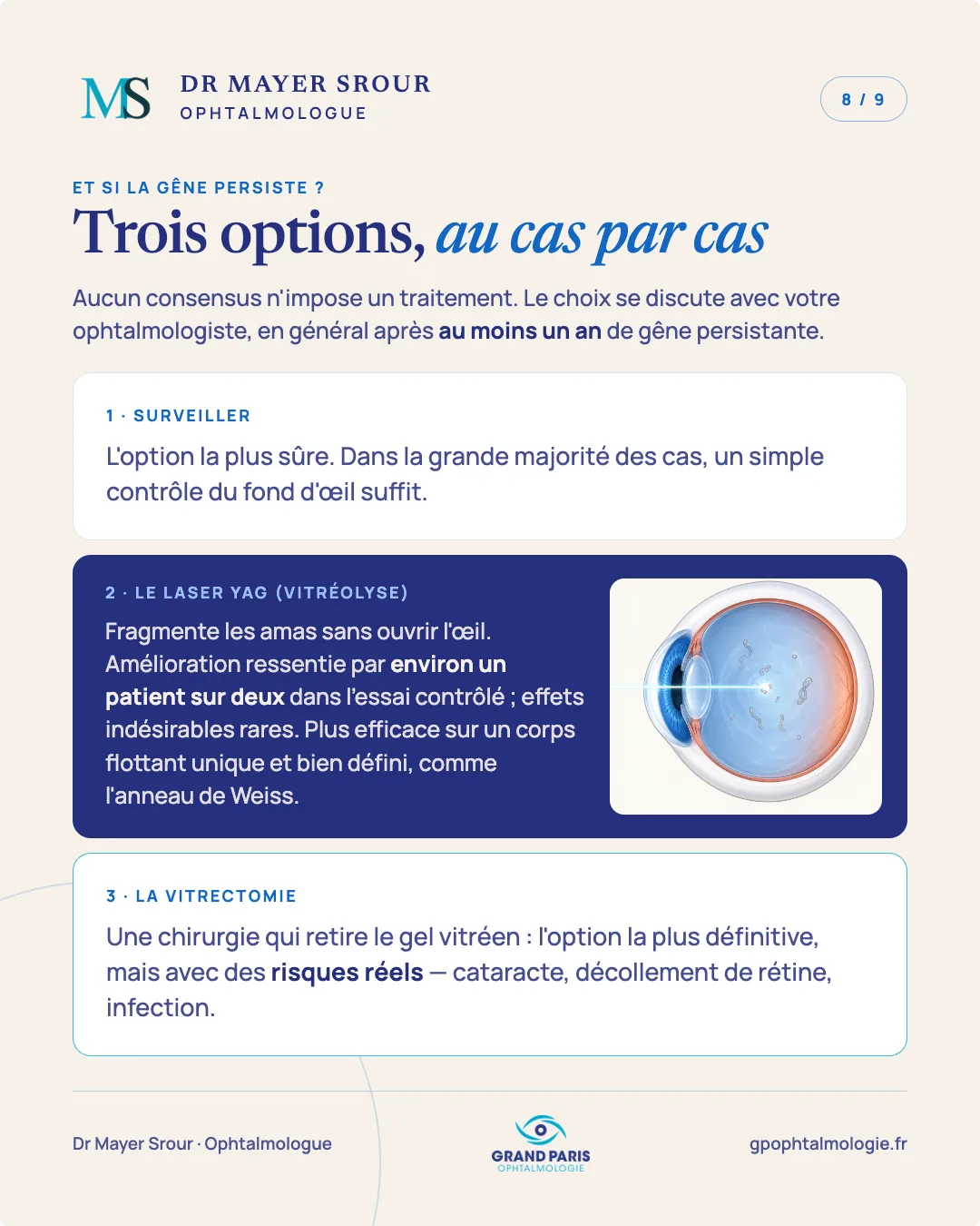
Three options, case by case
No consensus calls for treatment. The choice is discussed with your ophthalmologist, generally after at least a year of persistent trouble.
1 · Monitoring: the safest option. In the vast majority of cases, a simple fundus check is enough.
2 · YAG laser (vitreolysis): breaks up the clusters without opening the eye. Improvement felt by about one patient in two in the controlled trial; side effects are rare. More effective on a single, well-defined floater, such as the Weiss ring.
3 · Vitrectomy: surgery that removes the vitreous gel. The most definitive option, but with real risks — cataract, retinal detachment, infection.
What do we do at the practice?
A dilated fundus examination: drops dilate the pupil so the whole retina can be examined, right out to the periphery. Plan not to drive for 3 to 4 hours.
Key points: old, stable floaters are benign. New floaters, or floaters accompanied by flashes, should be examined without delay.
Sources: AAO Preferred Practice Pattern 2025 · Yang et al., Graefe's Archive 2023 · Alnajres et al., Ophthalmology Retina 2026 · Shah & Heier, JAMA Ophthalmology 2017 · Sebag, Prog Retin Eye Res 2020. For information only; does not replace a consultation.
Going further
This carousel summarises the essentials in a few images. The subject is covered in more depth on the dedicated page of the website: Floaters and flashes of light: when should you see a doctor?.