AMD: what a person with the condition may see
Age-related macular degeneration affects central vision after the age of 55. Signs, dry and wet forms, OCT assessment, prevention, injections and avenues of research: the carousel published on Instagram, in 6 images.
Swipe, or use the arrows. The text of each image is reproduced below.
The carousel, image by image
The text of each carousel page, to read at your leisure or to find a specific piece of information.
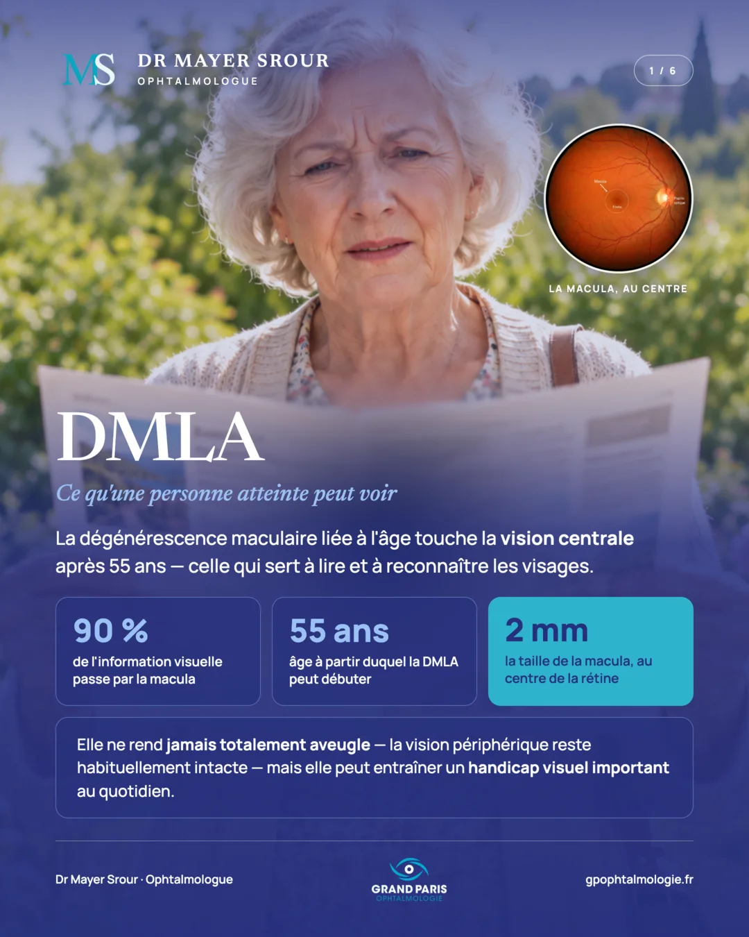
AMD: what a person with the condition may see
Age-related macular degeneration affects central vision after the age of 55 — the vision used for reading and recognising faces.
90% of visual information passes through the macula. 55 years: the age from which AMD can begin. 2 mm: the size of the macula, at the centre of the retina.
It never causes total blindness — peripheral vision usually remains intact — but it can lead to significant visual disability in everyday life.
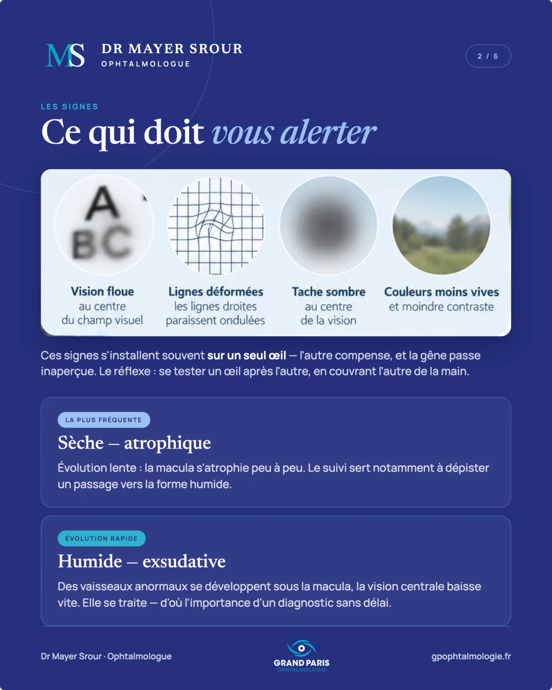
The signs: what should alert you
These signs often develop in one eye only — the other compensates, and the problem goes unnoticed. The reflex: test yourself one eye after the other, covering the other with your hand.
The most common — dry, atrophic: slow progression, the macula gradually wastes away. Follow-up is notably used to detect progression to the wet form.
Rapid progression — wet, exudative: abnormal vessels grow under the macula, and central vision falls quickly. It can be treated — hence the importance of a diagnosis without delay.
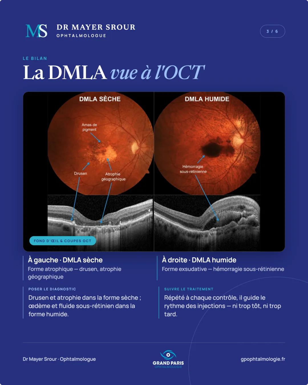
The assessment: AMD seen on OCT
Fundus photograph and OCT sections. On the left, dry AMD: atrophic form — drusen, geographic atrophy. On the right, wet AMD: exudative form — subretinal haemorrhage.
Making the diagnosis: drusen and atrophy in the dry form; oedema and subretinal fluid in the wet form.
Monitoring treatment: repeated at each check-up, OCT guides the pace of injections — neither too early nor too late.
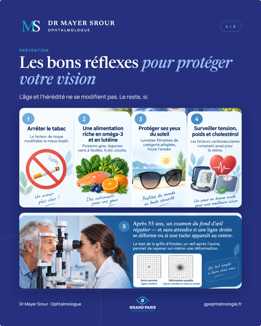
Prevention: the right reflexes to protect your vision
Age and heredity cannot be changed. The rest can: stopping smoking, a diet rich in leafy green vegetables and oily fish, sun protection, self-monitoring with the Amsler grid and regular follow-up.
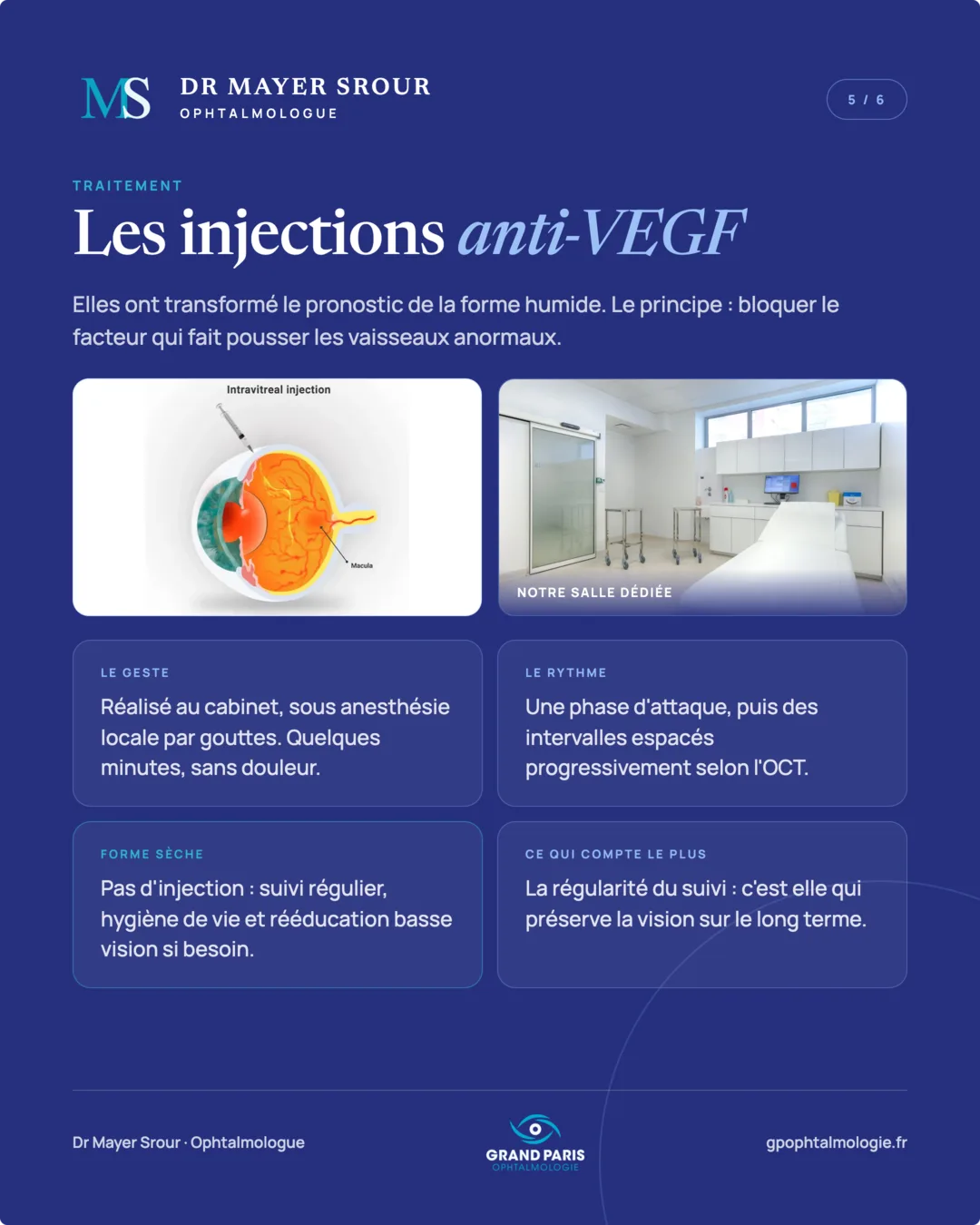
Treatment: anti-VEGF injections
They have transformed the outlook for the wet form. The principle: blocking the factor that makes abnormal vessels grow.
The procedure: performed at the practice, in our dedicated room, under local anaesthesia with drops. A few minutes, painless.
The schedule: a loading phase, then intervals gradually lengthened according to the OCT.
Dry form: no injection — regular follow-up, healthy lifestyle and low vision rehabilitation if needed.
What matters most: regular follow-up. This is what preserves vision in the long term.
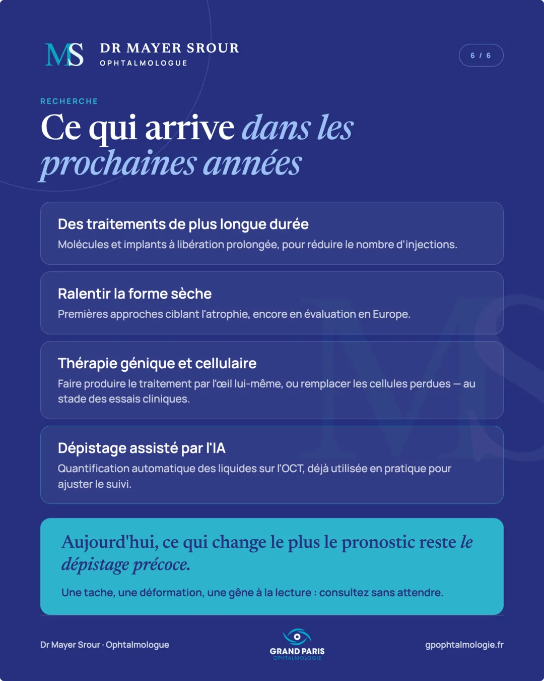
Research: what is coming in the next few years
Longer-lasting treatments: molecules and sustained-release implants, to reduce the number of injections.
Slowing the dry form: first approaches targeting atrophy, still under evaluation in Europe.
Gene and cell therapy: having the eye itself produce the treatment, or replacing lost cells — at the clinical trial stage.
AI-assisted screening: automatic quantification of fluid on OCT, already used in practice to adjust follow-up.
Today, what changes the outlook most remains early detection. A spot, a distortion, difficulty reading: see a doctor without delay.
Going further
This carousel summarises the essentials in a few images. The subject is covered in more depth on the dedicated page of the website: AMD: the practice's complete page.